Dr Ira Shah.
Medical Sciences Department, Pediatric Oncall, Mumbai, India. | | Case Report | A 2 1/2 months old male child born of 2nd degree consanguineous marriage presented with cough since 7 days, fever since 5 days and 5-6 episodes of generalized tonic clonic convulsions since 2 days. He was full term normal vertex vaginal hospital delivery without antenatal or post-natal complications and birth weight of 2.4 kgs. There was no history of rash or focal neurological deficits. On examination, he was febrile with a heart rate of 130/min. He had shallow breathing and anterior fontanelle was bulging. He had generalized lymphadenopathy (cervical, axillary and inguinal). His pupils were pinpoint and not reactive to light. On systemic examination, he had hypertonia with brisk reflexes and extensor planters. There were no meningeal signs. He had a moderate hepatosplenomegaly and on respiratory system examination had bilateral fine crepitations. In view of hepatosplenomegaly with generalized lymphadenopathy with raised intracranial tension and seizures, congenital infection was considered. His hemogram showed leucocytosis with anemia [Hb = 8.0 gm%, WBC = 28,200 cells/cu mm (51% polymorphs and 49% lymphocytes]. A CSF analysis was done which showed high proteins (147 mg%) with 4 polymorphs and 2 lymphocytes/cu mm and sugar of 45 mg/dl with corresponding blood sugar of 48 mg/dl. His liver function tests were normal and X-Ray chest showed interstitial pneumonia. TORCH titres were sent and his serum Toxoplasma IgG was positive [Toxo IgG = 170 IU/dl (Normal = <10 IU/dl)], and IgM Toxoplasma was negative. His CSF Toxoplasma IgG was 240 IU/dl suggestive of CNS Toxoplasmosis. His CMV, Rubella, Herpes simplex IgM & IgG were negative. His HIV by ELISA was negative. CT scan of the brain was done which showed extensive periependymal, subcortical white matter calcifications with basal and cisternal exudates and dilatation of right lateral ventricle suggestive of congenital intrauterine infection (Figure 1).
Figure 1 : CT scan showing extensive intracranial calcifications
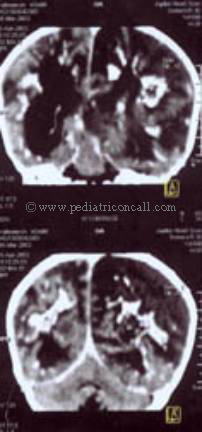
He was treated with spiramycin, pyrimethamine and Sulphadiazine. A chamber was inserted in the ventricular system and repeated chamber taps were required. He was also given blood transfusion for anemia. However, his clinical condition deteriorated and he finally succumbed to his disease within 3 days. Mother's TORCH titres were unavailable. | | | | Discussion | Toxoplasmosis is a zoonosis, the definitive host is the cat and all other hosts are incidental. Toxoplasma gondii is an obligate intracellular protozoan that can be acquired orally, transplacentally, parenterally by transfusion or from a transplanted organ. In normal children, infection may be asymptomatic, may lead to lymphadenopathy or damage any organ. Latent encysted organisms persist for life. Infections acquired congenitally almost always cause disease in perinatal period or later in life. Congenital transmission occurs when immunologically susceptible mother acquires infection during gestation. Chronic parasitemia inspite of presence of neutralizing antibodies in the mother may also lead to transplacental infection. (The possibility of parasitemia should be considered when a significant increase in IgG antibody titer occurs in a patient known to have a low stable titer of Toxoplasma antibodies.) Transmission may occur transplacentally or during labor. Of untreated maternal infections acquired in the first trimester, approximately 17% of fetuses are infected, usually with severe disease. Of untreated maternal infection acquired during 3rd trimester, approximately 65% of fetuses are infected usually with mild disease at birth. Transmission is less frequent when infection is acquired before 10th week of gestation and is exceptional when infection is acquired before conception.
Congenital infection during neonatal period may present as IUGR, prematurity, jaundice, thrombocytopenia, peripheral retinal scars, hepatosplenomegaly, microcephaly, convulsions, mental retardation, strabismus and cerebrospinal fluid pleocytosis along with the classical triad of chorioretinitis, hydrocephalus and cerebral calcifications. Infection may result in hydrops fetalis and even perinatal death. Sequelae are most often ocular. Ocular lesions may recur during childhood, adolescence or adulthood. In some instances, neurologic relapses have been observed.
Almost all organs of the body may be involved and manifestations may include. cutaneous rash, lymphadenopathy, myocarditis, pneumonitis, nephrotic syndrome, diarrhea, endocrine disorders and sensorineural deafness.
The diagnosis of acute infection with Toxoplasma may be established by isolation of T. gondii from blood or body fluids, demonstration of presence of cysts in placenta or tissues of a fetus or newborn, demonstration of presence of antigen and/or organisms in sections or preparations of tissues and body fluids, demonstration of antigenemia and antigen in serum and body fluids, PCR techniques or serologic tests.
Histologic diagnosis - Demonstration of tachyzoites in tissues (e.g., brain biopsy, bone marrow aspirate) or body fluids (cerebrospinal fluid, aqueous humor, sputum) establishes the diagnosis of toxoplasmosis.
Demonstration of antibodies in serum and body fluids - The most widely used serologic tests for the diagnosis of toxoplasma infection are Sabin-Feldman dye test, indirect hemagglutination (IHA) test, Immunofluroscence (IFA) test, Agglutination test, ELISA and ISAGA. The diagnosis of acute acquired toxoplasmosis may be established by the demonstration of rising serologic test titers.
The IHA test has frequently been negative in cases of congenital toxoplasmosis with high dye test titers and is therefore not recommended for the diagnosis of congenital toxoplasmosis. In addition because a rise in titer in the IHA test may not be demonstrable for months, it is not satisfactory as a screening method in pregnant women.
Complement fixing antibodies appear later than those demonstrable by the dye test. Thus CF test is useful in the demonstration of rising titers when dye test or IFA test titers are already high and stable. A negative CF test's turning positive or increasing CF test titers, together with stable high dye test titers indicate active infection. Sabin-Feldman dye test primarily measures IgG antibodies. It is sensitive and specific. IgG indirect fluorescent-antibody (IgG-IFA) test measures the same antibodies as the dye test and times tend to be parallel. IFA kits for T. gondii have been found to be improperly standardized. The IgM indirect fluorescent antibody (IgM-IFA) test is useful to diagnose acute infection in older children. It detects Toxoplasma-specific IgM in only approximately 25% of congenitally infected infants at birth. The IgM double sandwich ELISA detects approximately 75% of infants with congenital infection.
Serologic Diagnosis of Acquired Toxoplasma infection in pregnant woman: A rising titer is diagnostic. However, serologic test titers (e.g., dye or IFA) may have already reached their peak at the time the first serum is obtained for testing. Any patient with dye test or IFA test titers of higher than 300 IU/ml or 1:1000 and an IgM IFA test titer of 1:80 or higher or a capture IgM ELISA titer of 2 or higher must be presumed to have recently acquired infection with Toxoplasma until proved otherwise.
Serologic diagnosis is in the newborn: Persistent or rising titers in the dye or IFA test or a positive IgM ELISA is diagnostic of congenital toxoplasmosis. Presence of Toxoplasma-specific IgM in CSF that is not contaminated with blood or local antibody production of Toxoplasma specific IgG antibody demonstrated in CSF establishes the diagnosis of congenital toxoplasmosis.
Treatment
Pyrimethamine plus sulfadiazine are effective against toxoplasma. Use of pyrimethamine is contraindicated during 1st trimester of pregnancy. spiramycin should be used to prevent transmission of infection to the fetus of acutely infected pregnant women and to treat congenital toxoplasmosis.
All infected newborns should be treated whether or not they have clinical manifestations of the infection. Infants should be treated for 1 year with oral pyrimethamine (2 mg/kg/day for 2 days, then 1 mg/kg/day for 2-6 months, then 1 mg/kg/alternate day), sulfadiazine (100 mg/kg/day in divided doses) and leucovorin (5-10 mg/kg/day on alternate day). prednisone (1 mg/kg/day) can be used when active chorioretinitis involves the macula or otherwise threatens vision or CSF protein is > 1gm/dl.
Pregnant mothers with acute infection are treated with spiramycin (1 gm every 8 hours given without food) in 1st trimester. Ultrasonography and amniocentesis for PCR is done at 18 wk gestation. Fetal infection is treated with pyrimethamine and sulfadiazine or by termination of pregnancy.
Prognosis
Early therapy of congenital toxoplasmosis cures the manifestations. Sometimes hydrocephalus due to aqueductal obstruction may become worse on therapy. Children with extensive involvement at birth may function normally later in life or have mild vision, hearing, cognitive and neurologic. | | | | Compliance with Ethical Standards | | Funding None | | | | Conflict of Interest None | | |
- Behrman RE, Kliegman RM, Jenson HB - Nelson Textbook of Pediatrics, 17th ed. Philadelphia, W.B. Saunders, 2004: pg 1144-1153.
- Remington JS, Klein JO - Infectious Disease of the Fetus & Newborn Infant. Philadelphia, 4th ed, W.B. Saunders. 1995:pg 140-267.
|
| Cite this article as: | | Shah I. CONGENITAL TOXOPLASMOSIS. Pediatr Oncall J. 2004;1. |
|