Holly Paugh1, Yebabe Mengesha2.
1Tucson Hospitals Medical Education Program,
2Department of Pediatric Dermatology, Phoenic Childrens Hospital.
ADDRESS FOR CORRESPONDENCE
Holly Paugh, 3952 Paseo de las Canchas Tucson, AZ 85716, USA.
Email: hcpaugh@gmail.com | | Introduction | | Infantile myofibromas are the most common fibrous tumors of infancy. The clinical spectrum of infantile myofibromatosis is characterized by tumors in the skin, muscle, viscera, bone, and subcutaneous tissue. We present a case of an infant who presented with multiple congenital nodules who was diagnosed with infantile myofibromas. | | | | Case Report | A six-week-old male patient presented for evaluation of skin lesions, which were present since birth. The child also had a third lesion on the left forearm, which spontaneously regressed several weeks before his appointment. The patient's mother was concerned that the lesion on the left hand was causing the patient discomfort.
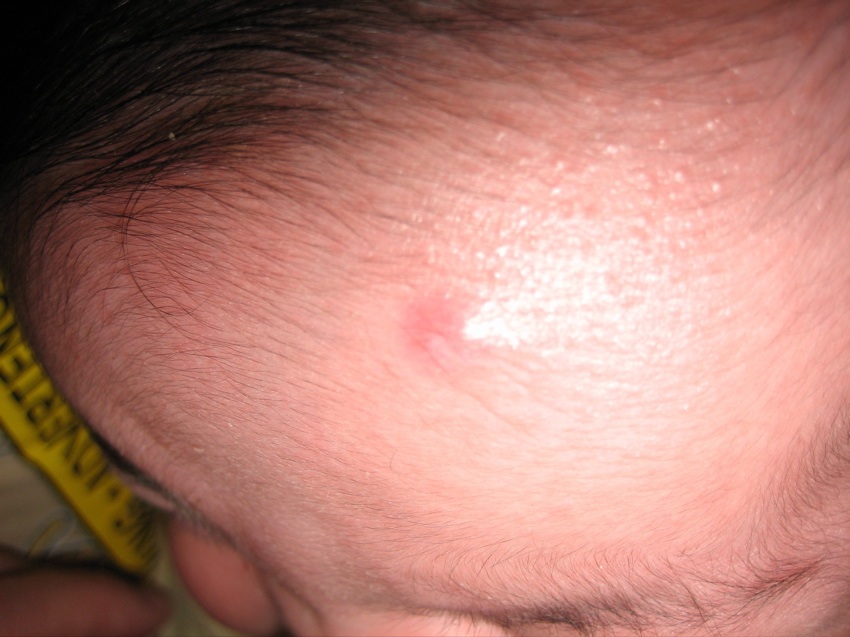
Physical examination revealed two skin lesions, a 1.5 cm subcutaneous nodular plaque in the first web space of the left hand {Figure 1} and a 6 mm dome shaped pink papule on the left forehead {Figure2}. The lesion was firm, tender and non-mobile. A 3 mm punch biopsy specimen was obtained from the lesion on the left hand for microscopic evaluation. Histopathology from the lesion on the left hand revealed fascicles resembling smooth muscle associated with thin spindle shaped nuclei with pointed ends, surrounded by abundant mucin. Chest and hand radiographs were ordered which showed no abnormalities. A diagnosis of infantile myofibromatosis was made based on the clinical and histologic findings. The clinical presentation of this patient with two left-sided lesions suggests a multicentric form of infantile myofibromatosis without visceral involvement. There was some concern that the lesion on the left hand could cause discomfort and disruption in fine motor skills. Given the likelihood of spontaneous resolution with time, clinical observation with follow-up was advised. At age 6 months, the lesion on the forehead and left hand were stable with no significant changes.
Figure 1: Nodular plaque on left hand
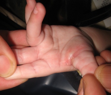
Figure 2: Nodular plaque on left Forehead
 | | | | Discussion | Three forms of infantile myofibromatosis have been described including 1) solitary infantile, 2) multicentric infantile without visceral involvement and 3) generalized infantile with visceral involvement. [7] The disorder was first described by Stout et al in 1954 under the term congenital generalized fibromatosis. [3] In 1981, Chung and Enzinger suggested changing the name to infantile myofibromatosis. [3]
Clinically, lesions appear as firm discrete nodules, ranging in size from 0.5-4.0 cm in diameter. [1,4,5] Solitary lesions are most often found in the head, neck, and shoulder girdle and may rapidly enlarge in the first few weeks of infancy. The clinical differential diagnosis of myofibromas involving soft tissue includes leiomyomas, hyaline fibromatosis, and soft tissue sarcomas. Biopsy is necessary for definitive diagnosis.
Histopathologic examination characteristically reveals a biphasic pattern composed of well-circumscribed but unencapsulated nodules of myofibrocytes resembling leiomyomas surrounding more cellular central areas resembling a hemangiopericytoma. [1,5] The myofibroblasts are spindle shaped and have tapering nuclei. Myxoid and pseudochondroid areas may also be seen. [1]
The prognosis of infantile myofibromatosis is dependent on the location and the number of lesions involved. Solitary tumors frequently involute spontaneously; whereas, the multicentric form with visceral involvement is usually associated with a poor prognosis. [1, 6, 7] Cardiopulmonary or gastrointestinal complications are the main causes of mortality often due to mass effect interfering with vital organ function. Therapeutic intervention and surgical treatment may be necessary if the disease is life threatening. [5]
| | | | Compliance with Ethical Standards | | Funding None | | | | Conflict of Interest None | | |
- Dimson, O.G., Drolet, B.A. et.al. Congenital generalized myofibromatosis in a neonate. Archives of Dermatology 2000; 136: 597-600. [CrossRef] [PubMed]
- Inamadar, A.C., Palit, A. et al Infantile Myofibromatosis with multiple congenital anomalies. Pediatric Dermatology. 2005;22:3. [CrossRef]
- Stout, Arthur, P. Juvenile Fibromatosis Cancer Sep 1954.
- Clarke, J.T., Clarke, L.E, et al. Plaque-like myofibroblastic tumor of infancy. Pediatric Dermatology 2007; E83-87. [CrossRef] [PubMed]
- Wiswell, T.E., Sakas, L., Stephenson, S. Infantile Myofibromatosis. Pediatrics. 1985. 76;6:981-984. [PubMed]
- Leaute-Labreze, C., Labarthe, M.P. et al. Self-Healing generalized infantile myofibromatosis with elevated urinary bFGF. Pediatric Dermatology. 2001;18:4 305-307.
- Variend, S. N.M.A. BAX, et al. Are infantile myofibromatosis, congenital fibrosarcoma and congenital haemangiopericytoma histogenetically related Histopathology 1995;26:57-62. [CrossRef]
|
| Cite this article as: | | Paugh H, Mengesha Y. Infantile Myofibromatosis. Pediatr Oncall J. 2008;5: 137-138. |
|