Satoshi Sato, Yasuyo Kashiwagi, Hisashi Kawashima, Tasuku Miyajima, Kouji Takekuma, Akinori Hoshika.
Department of Pediatrics, Tokyo Medical University, Japan.
ADDRESS FOR CORRESPONDENCE
Satoshi Sato, Tokyo Medical University, 6-7-1 Nishishinjuku, Shinjuku-ku, Tokyo 160-0023, Japan.
Email: satoshi115@nifty.com | An 11-month-old girl who refused to sit and showed increased irritability was admitted to Tokyo Medical University Hospital. She did not have any medical history. Ultrasound scan of the hips was normal. Her white cell count was 15,000/dl. C-reactive protein was 2.5 mg/dl. Her blood culture was sterile. Magnetic resonance imaging (MRI) confirmed the narrowing of an intervertebral disc space at L5-S1 (Fig 1a). Helical computed tomography (CT) scanning and reformation in sagittal image showed bone erosion (Fig 1b). These findings indicated spondylodiscitis. She responded well to antibiotics. The inflammatory markers had normalized and she was able to stand unsupported.
Fig 1a: Sagittal T2-weighted MRI spine
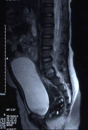
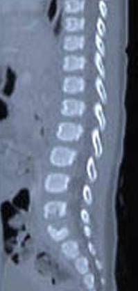
Spondylodiscitis (1, 2) is uncommon in infants. Clinical manifestations vary with age. Infants may present with irritability, toddlers may refuse to walk. MRI of the spine is diagnostic. It shows irregularity and destruction of the vertebral end plates and body, increased signal intensity on T2-weighted images, and bone destruction within adjacent vertebrae. Edema and purulent material in the marrow or disk space will appear as a dark signal on T1-weighted images and as a bright signal on T2-weighted images. CT was useful in providing a specific diagnosis of bone structure. CT scans also can identify the presence of adjacent soft tissue masses or abscesses. However, axial CT may miss destructive changes in spondylodiscitis. Reformatted CT sagittal images disclose spine damage much more obviously than axial slices alone.
Even though it is uncommon, the index of suspicion should be high in a child presenting with fever and refusal to sit or walk. | | | | Compliance with Ethical Standards | | Funding None | | | | Conflict of Interest None | | |
- An HS, Seldomridge JA. Spinal Infections: Diagnostic Tests and Imaging Studies. Clin Orthop Relat Res. 2006;444:27-33. [CrossRef] [PubMed]
- Kayser R, Mahlfeld K, Greulich M, Grasshoff H. Spondylodiscitis in Childhood: Results of a Long-Term Study. Spine. 2005;30:318-23. [CrossRef] [PubMed]
|
| Cite this article as: | | Sato S, Kashiwagi Y, Kawashima H, Miyajima T, Takekuma K, Hoshika A. Diagnosis of Spondylodiscitis by Magnetic Resonance Imaging and Computed Tomography. Pediatr Oncall J. 2008;5: 79. |
|