This case report details a 4-year-old previous healthy male who presented to the emergency department after experiencing fever for the past 4 days. In addition to the fever, he had a history of a productive cough that had progressively turned into a dry cough, along with a runny nose, over the course of the preceding week. Despite the absence of any signs of respiratory distress, the child’s mother was concerned about his persistent symptoms and the duration of the fever.
Upon clinical examination, the child appeared to be in overall good health. He was afebrile, displaying no signs of respiratory distress and no abnormalities in lung auscultation. The remaining examination was unremarkable.
A chest X-ray (CXR) was requested to evaluate for any potential underlying lung infection (Figure 1). The CXR showed no signs of lung consolidation or infiltrates. However, a hypotransparent, nodular lesion (6x4 mm) was observed in the right superior lobe linked to a fine convex line suggesting the possibility of an artifact, leading to a repeat CXR. Upon repeating the CXR, the lesion remained unchanged, which raised concerns. The radiographic finding was initially suspected to be a nodule or mass, prompting further investigation. To better characterize the lesion and determine its etiology, a thoracic CT scan was performed. The CT scan revealed a prominent accessory fissure associated with the azygos vein (Figure 2).
Figure 1. Chest X-ray showing accessory azygos fissure (AF), azygos lobe (AL) and azygos vein (AV).
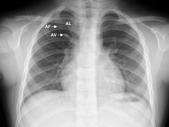 Figure 2.
CT scan showing accessory azygos fissure (AF), azygos vein (AV) and azygos lobe (AL).
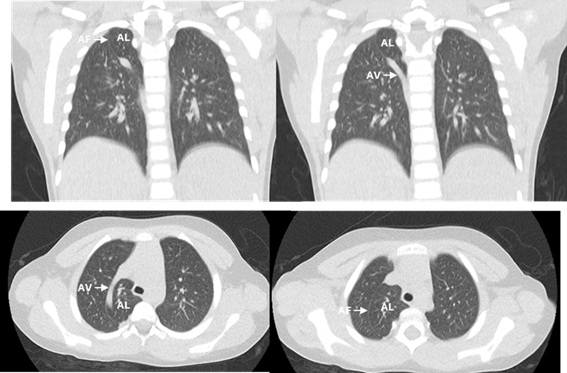
This finding was determined to be a normal anatomical variant, rather than a pathological nodule or mass. No other abnormalities were noted on the CT scan, including no signs of consolidation, pleural effusion, pericardial effusion, or lymphadenopathy. The findings confirmed the benign nature of the lesion and excluded more serious pulmonary conditions.
How can the azygos lobe be identified on a chest X-ray or CT scan?