|
Eye Lesion and Tuberculosis
Indrani Mondal, Nishit Anand, Pooja Mishra, Santosh Kumar.
Department of Pediatrics, Mata Gujari Memorial (MGM) Medical College and Lions Seva Kendra Hospital (LSKH), Kishanganj, Bihar, India.
ADDRESS FOR CORRESPONDENCE
Dr Santosh Kumar, F-110, New Doctors Hostel, MGM campus, Kishanganj, Bihar, India.
Email: santoshaiims08@gmail.com
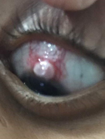
Figure 1: Greyish white nodular lesion along upper limbal region in left eye

|
|
What is the eye lesion?
Phlyctenular keratoconjunctivitis (PKC). It is a type IV cell mediated hypersensitivity reaction to endogenous microbial proteins in the cornea and/or conjunctiva, to a variety of antigens and is mostly unilateral with a strong female preponderance. (1-3) It is relatively more common in the pediatric age group in association with pulmonary and lymph node tuberculosis. (1) Many antigens like staphylococcal products, worm infestation, fungi, viruses, and parasites can cause it but the main antigen responsible for PKC is tuberculoprotein. (2,3) PKC is a nodular affliction characterized by the formation of a small circumscribed lesions at the corneal limbus. (4) Conjunctival phlyctens are usually transient and asymptomatic but corneal lesions present with lacrimation, photophobia and blepharospasm and may leave residual opacities leading to permanent vision impairment. (5) PKC may be a presenting feature in a patient without any systemic tuberculosis symptoms. (3) PKC lesions are observed to be more severe and recurrent in patients with tuberculosis especially with pulmonary tuberculosis and tubercular lymphadenopathy. (5) Topical corticosteroids and local antibiotics are considered as the best options for PKC. A routine ophthalmological evaluation in all patients with tuberculosis will help in early diagnosis and timely institution of local treatment of PKC. |
| |
| Compliance with ethical standards |
|
Funding: None
|
|
|
Conflict of Interest: None
|
|
- Bhandari A, Bhandari H, Shukla R, Giri P. Phlyctenular conjunctivitis: a rare association with spinal intramedullary tuberculoma. BMJ Case Rep 2014 Mar 18; pii: bcr2013202010. [CrossRef]
- Rohatgi J, Dhaliwal U. Phlyctenular eye disease: A reappraisal. Jpn J Ophthalmol. 2000;44:146–50. [CrossRef]
- Valentina C, Mirela C. Phlyctenular keratoconjunctivitis and lymph node tuberculosis. Oftalmologia. 1999;48:15-8 [PubMed]
- Gautam P, Shrestha GS, Sharma AK. Phlyctenular keratoconjunctivitis among children in the tertiary eye hospital of Kathmandu, Nepal. Oman Journal of Ophthalmology. 2015;8:147-150. [CrossRef] [PubMed] [PMC free article]
- Singal A, Aggrawal P, Pandhi D, Rohatgi J. Cutaneous tuberculosis and phlyctenular keratoconjunctivitis: A forgotten association. Indian J Dermatol Venereol Leprol 2006;72:290-2. [CrossRef] [PubMed]
|
|
DOI: https://doi.org/10.7199/ped.oncall.2018.19 |
| |
Cite this article as:
Mondal I, Anand N, Mishra P, Kumar S. Eye Lesion and Tuberculosis. Pediatr Oncall J. 2018;15: 50. doi: 10.7199/ped.oncall.2018.19
|