|
Bone Marrow Abnormality in a case of Chronic Myeloid Leukemia
Govind Kendre, Shruti Mantri, Sunil Hilalpure, Suraj Goyanka, Leo Prince, Murlidharan C, Chandrakala S, Farah Jijina.
Department of Clinical Hematology, Seth G.S. Medical College, KEM Hospital, Mumbai, India.
ADDRESS FOR CORRESPONDENCE
Dr. Govind Kendre, Department of Clinical Hematology, Seth G.S. Medical College, KEM Hospital, Parel, Mumbai.
Email: govindken143@gmail.com
CML, sea-blue-histiocyte, bone marrow
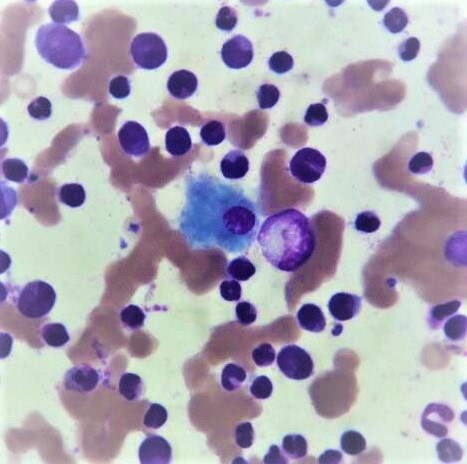
An 11-year-old boy presented with fatigue for 4 months. There was no fever, breathlessness, weight loss, paleness, jaundice or bleeding from any site. On physical examination spleen was palpable 10 cm below left costal margin along its long axis. No other significant abnormality was detected. Complete blood count (CBC) revealed hemoglobin 10.5 g/dL, platelet count 179,000 cells/cumm, and white blood cell count 64,000 cells/cumm. Peripheral blood smear showed marked leukocytosis with left shift of granulocytes. Bone marrow smear examination showed hypercellular marrow and hypo-lobulated megakaryocytes with a differential count of erythroid precursors (10%), myelocytes and metamyelocytes (30%), band cells (30%), mature neutrophils (20%), myeloblasts (5%) and lymphocytes (5%). In the background of increased myeloid precursors, prominent macrophages were seen, the cytoplasm of which was closely packed with fine granules staining blue with Giemsa and the nuclei was displaced towards the periphery (Figure 1). Further investigations showed the presence of BCR-ABL1 fusion by fluorescent in situ hybridization, as well as by PCR, thus confirming the diagnosis of chronic myelogenous leukemia (CML).
Figure 1. Bone marrow aspirate smear (Giemsa stain) (1000x)
|
What is the bone marrow aspirate smear suggestive of?
The bone marrow aspirate smear is suggestive of sea-blue-histiocyte. The sea-blue histiocytes are macrophages, varying from 20 to 60 um in diameter with a single eccentric nucleus, large cytoplasm, and sea-blue granules containing lipofuscin or ceroid.1 Sea blue histiocytes are a common feature of CML and that their accumulation seems to be associated with a prolonged increase in leucocyte turnover thus, pointing towards the chronic phase of CML, although this finding has no prognostic importance in the era of tyrosine kinase inhibitors (TKI). Besides CML, sea-blue–histiocytes have also been described in other conditions such as myelodysplastic syndromes, lymphomas, idiopathic thrombocytopenic purpura, lipid storage diseases and ß-thalassemia major.2
Acknowledgment
We thank Dr. Chandrakala S (HOD clinical hematology) and Dr. Farah Jijina for their valuable inputs. |
Cite this article as:
Kendre G, Mantri S, Hilalpure S, Goyanka S, Prince L, C M, S C, Jijina F. Bone Marrow Abnormality in a case of Chronic Myeloid Leukemia. Pediatr Oncall J. 2020;17: 27. doi: 10.7199/ped.oncall.2020.8
|