|
Aicardi-Goutières Syndrome - a neonatal clinical report
Bárbara Ribeiro de Aguiar1, Sara Torres Oliveira2, Hugo Cavaco3, Sara Noéme Prado3, Joana Saldanha3, Sofia Quintas4.
1Department of Pediatrics, Hospital Beatriz Ângelo, Loures, Portugal,
2Department of Pediatrics, Hospital de Santa Maria, University Hospital Center of North Lisbon, Lisbon, Portugal,
3Neonatal Intensive Care Unit, Department of Pediatrics, Hospital Beatriz Ângelo, Loures, Portugal,
4Neuropediatrics Unit, Department of Pediatrics, Hospital de Santa Maria, University Hospital Center of North Lisbon, Lisbon, Portugal.
ADDRESS FOR CORRESPONDENCE
Bárbara de Paiva Morão Ribeiro de Aguiar, Pediatric Department, Hospital Beatriz Ângelo, 2674-514 Loures, Portugal.
Email: b.ribeiroaguiar@gmail.com
Aicardi-Goutières syndrome, Neonate, Intracerebral calcifications, Basal ganglia calcifications.
We present a case involving a male newborn born to healthy parents after a pregnancy with appropriate surveillance and no complications. Maternal serologic screening and blood tests showed normal results. The membranes spontaneously ruptured two hours before delivery, with clear amniotic fluid. The baby was delivered vaginally at 40 weeks gestational age, with Apgar scores of 9 at 1st minute and 10 at 5th minute. Birth measurements were within the 50th, 3rd, and 15th percentiles for weight, height, and head circumference, respectively.
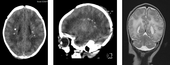
On the second day of life, multiple petechiae were noted, and hematological examinations disclosed thrombocytopenia with a count of 63 · 10^9 cells/L. Head ultrasonography indicated the presence of calcifications in the thalamic and periventricular regions (Figure 1). Due to suspected congenital TORCH infection, the newborn was transferred to the Neonatal Intensive Care Unit. Imaging studies showed abnormalities in cerebral white matter, subcortical atrophy, and calcifications around the basal ganglia and periventricular regions (Figure 2). The newborn underwent various tests, excluding intrauterine congenital infections, as well as disorders of calcium and phosphorus metabolism, cerebral folate deficiency and mitochondrial diseases. The results of the electroencephalogram showed no abnormalities. Ophthalmological, neurologic, and cardiac assessments yielded normal results.
Figure 1. Head ultrasonography suggested thalamic and periventricular calcifications.
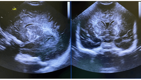
Figure 2. Computed tomography and magnetic resonance imaging of the brain showing attenuation of cerebral white matter, subcortical atrophy and basal ganglia and periventricular calcifications.
By the third week, thrombocytopenia had resolved, and the newborn was released for additional assessments in Pediatric Neurology, specifically involving genetic analysis and cerebrospinal fluid.
During the initial months of life, a mild lag in psychomotor development became apparent, leading to the performance of a lumbar puncture. At the four-month follow-up, findings included gastroesophageal reflux and feeding discomfort, notable delays in psychomotor developmental, spastic-dystonic tetraparesis, irritability and developed microcephaly characterized by a head circumference below the 3rd percentile. A follow-up electroencephalogram displayed a widespread slowing pattern. Cerebrospinal fluid analysis identified lymphocytosis, elevated interferon alpha, and increased neopterin levels. Consequently, the patient commenced pulses of methylprednisolone. The genetic examination unveiled a homozygous mutation in the TREX1 gene, leading to the diagnosis of Aicardi-Goutières syndrome. Treatment with a JAK inhibitor (baricitinib) was initiated at nine months, resulting in clinical stabilization and cessation of developmental regression. The patient acquired head control, started smiling, displayed good interaction and irritability disappeared. A Percutaneous Endoscopic Gastrostomy was placed, and a Nissen fundoplication was performed, resulting in an improvement in gastroesophageal reflux.
|
What is the diagnosis?
Aicardi-Goutières syndrome represents an uncommon genetic neurological condition characterized by an overproduction of interferon, leading to systemic inflammatory damage.1,2,3 This syndrome is marked by non-specific neurological symptoms and systemic manifestations, often linked to intracerebral calcifications, white matter abnormalities, cerebral atrophy, cerebrospinal fluid lymphocytosis, and elevated interferon alpha levels.3,4,5 Due to the resemblance of its neurological symptoms to other disorders, diagnosing the syndrome is quite challenging.6,7,8 Neonatal onset of this condition is exceedingly rare and is most frequently associated with the TREX gene.9 This case underscores the importance of considering Aicardi-Goutières syndrome as a potential differential diagnosis in newborns presenting intracerebral and basal ganglia calcifications, particularly after excluding infections, calcium and phosphorus metabolism disorders, cerebral folate deficiency, and mitochondrial diseases. |
- Volpe, Joseph J. Neurology of the newborn, 5th edition (2008), 730.
- Adang L, Gavazzi F, Simone M, et al. Developmental Outcomes of Aicardi Goutières Syndrome. Journal of Child Neurology 2020; 35(1):7-16. [CrossRef] [PubMed] [PMC free article]
- Crow Y. Aicardi-Goutières syndrome. Handbook of Clinical Neurology. Elsevier 2013; 113:1639-35. [CrossRef] [PubMed]
- Tonduti D, Fazzi E, Badolato R, et al. Novel and emerging treatments for Aicardi-Goutières syndrome. Expert Review of Clinical Immunology 2020; 16(2):189-98. [CrossRef] [PubMed]
- Livingston JH, Stivaros S, Warren D, et al. Intracranial calcification in childhood: A review of aetiologies and recognizable phenotypes. Developmental Medicine and Child Neurology 2014; 56:612-26. [CrossRef] [PubMed]
- Rice G, Patrick T, Parmar R, et al. Clinical and Molecular Phenotype of Aicardi-Goutières Syndrome. The American Journal of Human Genetics 2007; 81:713-25. [CrossRef] [PubMed] [PMC free article]
- La Piana R, Uggetti C, Roncarolo F, et al. Neuroradiologic patterns and novel imaging findings in Aicardi-Goutieres syndrome. Neurology 2016; 86:28-35. [CrossRef] [PubMed] [PMC free article]
- Uggetti C, La Piana R, Orcesi S, et al. Aicardi-Goutieres syndrome: neuroradiologic findings and follow-up. NR Am J Neuroradiol 2009; 30(10):1971-6. [CrossRef] [PubMed] [PMC free article]
- Moreno Medinilla EE, Villagrán García M, Mora Ramírez MD, et al. Síndrome de Aicardi-Goutières: espectro fenotípico y genético en una serie de 3 casos. An Pediatr (Barc). 2019;90:312-314. [CrossRef] [PubMed]
|
|