|
An atypical cause of a neck mass
Daniela Araújo, Mariana Pereira, Joana Vilaça, Joana Oliveira, Ângela Oliveira, Helena Silva.
Pediatrics Department, Unidade Local de Saúde de Braga, Braga, Portugal.
ADDRESS FOR CORRESPONDENCE
Daniela Araújo, Sete Fontes - São Victor, 4710-243 Braga, Portugal.
Email: da.niielamatos@hotmail.com
parotid abscess, myositis, Bartonella henselae, complicated cat scratch disease
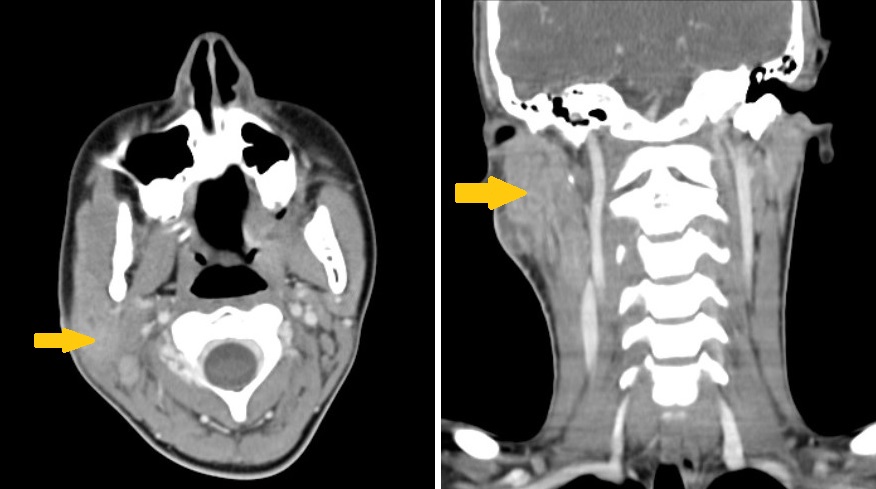
A previously healthy 13-year-old female presented to the Emergency Department (ED) with a painful right cervical mass that had persisted for approximately one month (Figure 1). Initial symptoms included mild odynophagia, which had resolved by the time of admission. The patient denied fever, night sweats, or significant exposure to known infections, although she lived in a rural area with daily contact with a cat and a dog. Her mother had regular contact with a tuberculosis patient at work.
Figure 1. Computerized tomography (CT) findings.
Two weeks prior, her family doctor had ordered blood tests and a cervical ultrasound, which revealed two nodular formations. Despite a 7-day course of oral Amoxicillin with Clavulanic Acid and a 5-day anti-inflammatory regimen, the mass continued to enlarge.
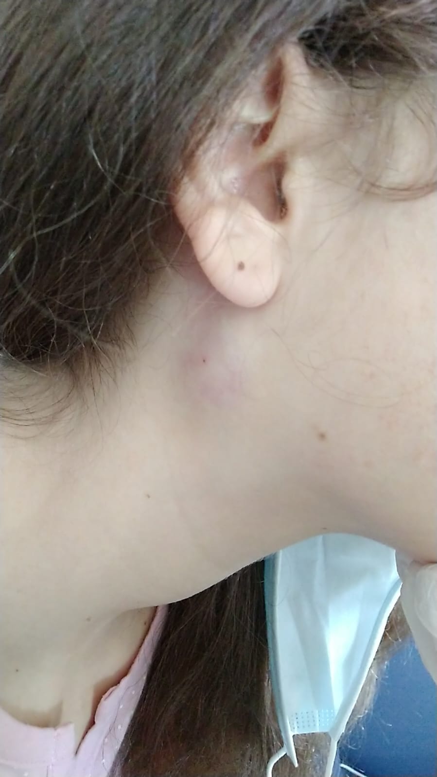
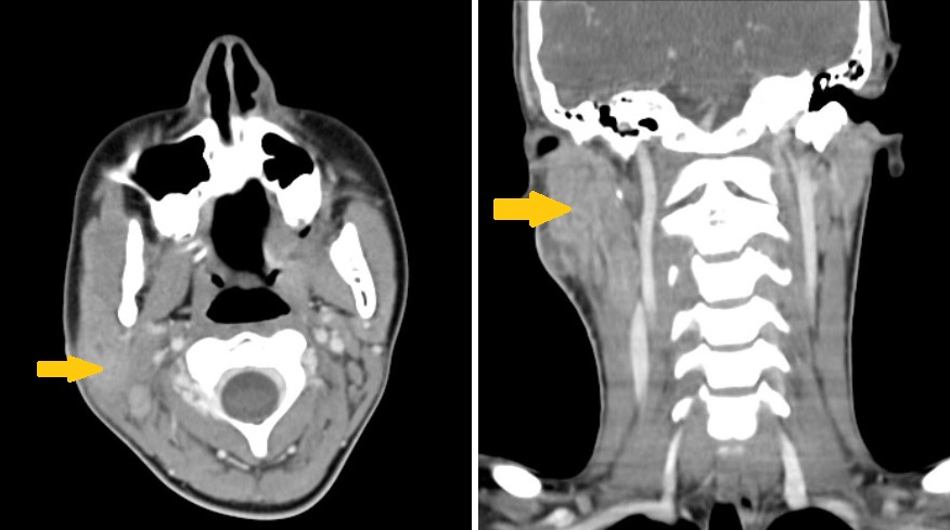
On admission to the ED, a physical exam revealed a 3 cm non-tender, painful cervical swelling with several smaller adjacent swellings. Initial lab results were within normal ranges, and CRP was mildly elevated of 2.1 mg/L (Table 1). A subsequent ultrasound revealed a hypoechogenic mass with irregular contours and surrounding fat hyperechogenicity, suggestive of abscess formation. A cervical CT confirmed a right parotid abscess and ipsilateral platysma myositis (Figure 2).
Table 1. Results from serological testing.
| Serological tests |
Findings |
| Anti-CMV |
Negative (IgG and IgM) |
| EBV early antigen |
Negative for IgG |
| EBV viral capsid antigen |
IgG positive; IgM negative |
| EBV nuclear antigen |
Negative for IgG |
| Herpes simplex 1 |
Negative (IgG and IgM) |
| Herpes simplex 2 |
Negative (IgG and IgM) |
Figure 2. Tumefactions inflammatory signs on the first day of hospitalization.
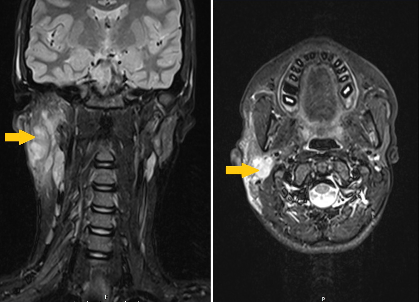
The patient was started on intravenous ceftriaxone, clindamycin, and oral azithromycin. Despite initial treatment, inflammatory signs worsened. Aspiration was performed and revealed purulent fluid, which was sent for microbiological analysis. MRI confirmed the abscess and surrounding cellulitis (Figure 3). After four days, the patient showed improvement, though the mass persisted, prompting a core biopsy.
Figure 3. Magnetic resonance imaging (MRI) findings.
|
Figure 1. Computerized tomography (CT) findings.
|
What can be the cause of this subacute lymphadenopathy?
On day 8, DNA testing of the purulent material confirmed Bartonella henselae and ruled out tuberculosis.
Lymphadenopathies are frequently encountered in the paediatric Emergency Department (ED). They can be categorized based on duration as acute (<3 weeks), subacute (3-6 weeks), or chronic (>6 weeks).1 The aetiology of lymphadenopathy encompasses inflammatory, benign noninflammatory, and malignant causes.2,3Among the various causes, viral infections represent the most common aetiology of cervical lymphadenopathy.1 However, bacterial infections can also be a contributing factor.1
The patient completed a 10-day intravenous antibiotic course and a 5-day course of oral azithromycin and was discharged. Follow-up at two months revealed complete resolution, with no palpable lymphadenopathy. Histology showed a non-specific granulomatous inflammatory process.
This case report highlights a rare and complicated presentation of Cat Scratch Disease (CSD) in an adolescent, characterized by cellulitis, myositis of the sternocleidomastoid muscle (SCM), and a parotid abscess. CSD is caused by Bartonella henselae, one of the three Bartonella species responsible for the majority of human infections.4,5 Domestic cats, particularly those under one year old, are the primary reservoirs for B. henselae.4,6 However, as demonstrated in our patient, a history of a definitive cat scratch or bite is not always evident, underscoring the need for a high index of clinical suspicion.4,6
In immunocompetent individuals, CSD typically presents as subacute, regional lymphadenopathy.5 The disease often begins with fever and the appearance of an erythematous papule at the site of inoculation.6 This is followed by lymphadenopathy in the draining lymph nodes, with cervical and axillary nodes being the most frequently affected.4,6Less commonly, nodes in the submandibular, preauricular, femoral, and inguinal regions may be involved, while parotid gland involvement, as in our case, is rare.7
The diagnosis of uncomplicated CSD is often clinical, based on characteristic symptoms and epidemiological history. Confirming Bartonella infection via culture is notoriously difficult, making serological testing the mainstay for identifying atypical presentations.8 However, serologic testing has its limitations, as positive results may reflect prior exposure rather than active infection.8
While most mild to moderate cases resolve spontaneously within one to two months without specific treatment, complicated CSD, as seen here, requires intervention.9 Antibiotic therapy is generally reserved for cases involving severe symptoms, significant morbidity, or atypical manifestations.9
This case is particularly noteworthy due to its unusual presentation. Despite extensive infection involving cellulitis, myositis, and abscess formation, the patient lacked fever and showed normal inflammatory markers, which are atypical for such severe involvement. These findings emphasize the importance of maintaining clinical suspicion for CSD, even in the absence of classic inflammatory signs.
Detailed epidemiological investigation, including pet exposure history, was pivotal in raising clinical suspicion in this case. When antibiotic treatment does not yield expected improvements, clinicians should consider potential complications such as abscess formation. Early recognition and intervention are essential in minimizing morbidity and achieving favourable outcomes. |
| |
| Compliance with ethical standards |
|
Funding: None
|
|
|
Conflict of Interest: None
|
|
- Weinstock MS, Patel NA, Smith LP. Pediatric cervical lymphadenopathy. Pediatr Rev. 2018;39(9):433-443. doi:10.1542/pir.2017-0249. [CrossRef] [PubMed]
- Eapen A, Kamat D. Neck masses in children. Clin Pediatr (Phila). 2014;53(11):1027-1029. doi:10.1177/0009922813512177. [CrossRef] [PubMed]
- Meier JD, Grimmer JF. Evaluation and management of neck masses in children. Am Fam Physician. 2014;89(5):353-358.
- English R. Cat-Scratch Disease. Pediatr Rev. 2006;27(4):123-128. [CrossRef] [PubMed]
- Prutsky G, Domecq JP, Mori L, et al. Treatment outcomes of human bartonellosis: A systematic review and meta-analysis. Int J Infect Dis. 2013;17(10):e811-e819. doi:10.1016/j.ijid.2013.02.016. [CrossRef] [PubMed]
- Nawrocki CC, Max RJ, Marzec NS, Nelson CA. Atypical manifestations of cat-scratch disease, United States, 2005-2014. Emerg Infect Dis. 2020;26(7):1438-1446. doi:10.3201/eid2607.200034. [CrossRef] [PubMed] [PMC free article]
- Petrogiannopoulos C, Valla K, Mikelis A, et al. Parotid mass due to cat scratch disease. Int J Clin Pract. 2006;60(12):1679-1680. doi:10.1111/j.1742-1241.2005.00657.x. [CrossRef] [PubMed]
- Klotz, S.A., Ianas, V., Elliott SP. Cat-scratch Disease. Am Fam Physician 153. 2011;83(2):152-155. https://pubmed.ncbi.nlm.nih.gov/21243990/.
- Zangwill KM. Cat Scratch Disease and Bartonellaceae: The Known, the Unknown and the Curious. Pediatr Infect Dis J. 2021;40(5):S11-S15. doi:10.1097/INF.0000000000002776. [CrossRef] [PubMed]
|
|
DOI: https://doi.org/10.7199/ped.oncall.2026.70 |
| |
Cite this article as:
Araújo D, Pereira M, Vilaça J, Oliveira Â, Silva H, Oliveira J. An atypical cause of a neck mass. Pediatr Oncall J. 2025 Jan 24. doi: 10.7199/ped.oncall.2026.70
|