|
Ischemic stroke in a 6 month old infant! What's behind?
Carolina Simão1, Inês Carvalho1, Sarah Stokreef1, Carla Conceição2.
1Pediatric Department, Hospital do Divino Espirito Santo de Ponta Delgada EPER, 9500-782 Ponta Delgada São Miguel Island, Azores, Portugal,
2Neuroradiology Department, Hospital Dona Estefânia, 1169-045, Lisbon, Portugal.
ADDRESS FOR CORRESPONDENCE
Carolina Simão, Rua do Seixal, n15, Netos, Ferreira a Nova, Figueira da Foz, Coimbra, Portugal.
Email: carolin.simao@hotmail.com
neurology, neurosurgery, moyamoya, stroke
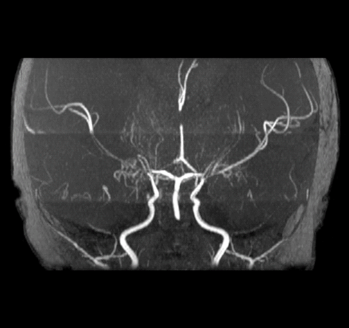
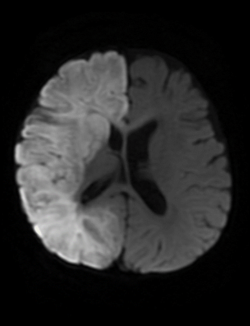
A 6-month-old infant was admitted to the Emergency Department due to prostration and focal seizures for the last 3 days. On admission he was hypotonic, had right side eye deviation and later developed left sided hemiparesis. Cranial CT showed an extensive heterogeneously hypodense lesional area involving the entire right cerebral hemisphere. (Figure 1) The electroencephalogram showed right temporal epileptic activity suggesting focal status epilepticus. Brain MRI revealed an extensive ischemic stroke in the right hemisphere involving the internal carotid artery (ICA) and posterior cerebral artery territories, as well as a small contralateral infarction in the middle cerebral artery/barrier territory. MR angiography showed a bilateral pre-obliterative stenosis of the distal ICA and proximal segments of the anterior and middle cerebral arteries with lenticulostriate branch hypertrophy. (Figure 2) The genetic study revealed a heterozygous variant in the RNF213 gene.
After revascularization surgery a partial focal deficit recovery was observed, with remnant moderate psychomotor development delay.
Figure 1. Cranial CT showed an extensive heterogeneously hypodense lesional area involving the entire right cerebral hemisphere.
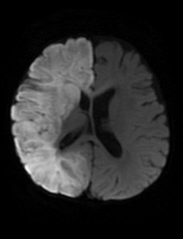
Figure 2 MR angiography showed a bilateral pre-obliterative stenosis of the distal ICA and proximal segments of the anterior and middle cerebral arteries.
|
Figure 1. Cranial CT showed an extensive heterogeneously hypodense lesional area involving the entire right cerebral hemisphere.
|
What is the diagnosis?
Moyamoya disease is a rare, idiopathic, progressive cerebrovascular disorder characterized by stenosis or occlusion of the terminal portion of the internal carotid arteries and their main branches, leading to the development of a network of fragile collateral vessels at the base of the brain. This network of vessels gives a characteristic “puff of smoke” appearance on angiography, which is the origin of the name “moyamoya”, a Japanese term meaning “something hazy like a puff of smoke”.1,2,3
The disease has a bimodal age distribution, with peaks in childhood (typically presenting with ischemic symptoms) and in adulthood (presenting with both ischemic and hemorrhagic symptoms).1,2,3 Moyamoya disease is more prevalent in East Asian populations, particularly in Japan, and has a higher incidence in females.1,3,4
The pathogenesis of moyamoya disease is not fully understood, but genetic factors, such as mutations in the RNF213 gene, have been implicated, particularly in East Asian populations.1,3,5 Moyamoya syndrome refers to similar vascular changes associated with other conditions like Down syndrome, sickle cell disease, and prior radiation exposure.2,6
The RNF213 gene, particularly the p.R4810K variant, has been identified as a major susceptibility gene for Moyamoya disease, especially in East Asian populations. This variant is associated with higher risk of early-onset disease, more severe clinical manifestations, and a higher likelihood of familial cases.7
Moyamoya’s CT angiography shows a stenosis or occlusion of the distal internal carotid arteries (ICAs) and their proximal branches, a collateral vessel formation at the base of the brain and dilatation of the anterior choroidal and posterior communicating arteries. An Magnetic Resonance Angiography shows absence of flow voids in the internal carotid arteries and prominent flow voids from basal ganglia and thalamic collateral vessels, indicating the presence of abnormal collateral circulation.
Management of Moyamoya disease focuses on preventing stroke and improving cerebral blood flow. Surgical revascularization is the primary treatment for symptomatic patients. Direct bypass procedures, such as superficial temporal artery to middle cerebral artery anastomosis, and indirect procedures, such as encephaloduroarteriosynangiosis, are commonly employed. The American Heart Association/ American Stroke Association guidelines recommend surgical intervention in symptomatic patients to prevent stroke recurrence.2 Antiplatelet therapy may be used as an adjuvant to prevent recurrent ischemic events.1
Moyamoya disease are associated with high morbidity. Early diagnosis and proper treatment are crucial to minimize neurological sequelae. |
| |
| Compliance with ethical standards |
|
Funding: None
|
|
|
Conflict of Interest: None
|
|
- Kappel AD, Feroze AH, Torio E, Sukumaran M, Du R. Management of moyamoya disease: a review of current and future therapeutic strategies. J Neurosurg. 2024;141(4):975-982. Published 2024 Apr 19. doi:10.3171/2024.1.JNS221977. [CrossRef] [PubMed]
- Kleindorfer DO, Towfighi A, Chaturvedi S, et al. 2021 Guideline for the Prevention of Stroke in Patients With Stroke and Transient Ischemic Attack: A Guideline From the American Heart Association/American Stroke Association [published correction appears in Stroke. 2021 Jul;52(7):e483-e484. doi: 10.1161/STR.0000000000000383.]. Stroke. 2021;52(7):e364-e467. doi:10.1161/STR.0000000000000375. [CrossRef]
- Kuroda S, Houkin K. Moyamoya disease: current concepts and future perspectives. Lancet Neurol. 2008;7(11):1056-1066. doi:10.1016/S1474-4422(08)70240-0. [CrossRef] [PubMed]
- Burke GM, Burke AM, Sherma AK, Hurley MC, Batjer HH, Bendok BR. Moyamoya disease: a summary. Neurosurg Focus. 2009;26(4):E11. doi:10.3171/2009.1.FOCUS08310. [CrossRef] [PubMed]
- Ihara M, Yamamoto Y, Hattori Y, et al. Moyamoya disease: diagnosis and interventions. Lancet Neurol. 2022;21(8):747-758. doi:10.1016/S1474-4422(22)00165-X. [CrossRef] [PubMed]
- Dlamini N, Muthusami P, Amlie-Lefond C. Childhood Moyamoya: Looking Back to the Future. Pediatr Neurol. 2019;91:11-19. doi:10.1016/j.pediatrneurol.2018.10.006. [CrossRef] [PubMed]
- Shlobin NA, Hoffman SC, Clark JR, Du RY, Lam S. Clinical Usefulness of Genetic Testing For Patients with Moyamoya Disease: A Systematic Review. World Neurosurg. 2021;152:198-205.e1. doi:10.1016/j.wneu.2021.05.130. [CrossRef] [PubMed]
|
|
DOI: https://doi.org/10.7199/ped.oncall.2026.72 |
| |
Cite this article as:
Simão C, Carvalho I, Stokreef S, Conceição C. Ischemic stroke in a 6 month old infant! What's behind?. Pediatr Oncall J. 2025 Mar 07. doi: 10.7199/ped.oncall.2026.72
|