|
Azygos Accessory Fissure in a Pediatric Patient
Ana Sofia dos Santos Lopes da Silva1, Madalena Torrado Malato2, Daniela Figueiredo Ramos2.
1Unidade Local de Saúde de Santa Maria, Hospital de Santa Maria, Lisboa, Portugal,
2Unidade Local de Saúde Estuário do Tejo, Hospital de Vila Franca de Xira, Vila Franca de Xira, Portugal.
ADDRESS FOR CORRESPONDENCE
Ana Sofia dos Santos Lopes da Silva, Estrada Carlos Lima Costa Nº2, Povos, 2600-009 Vila Franca de Xira.
Email: anasofiaslsilva1@gmail.com
Azygos accessory fissure, Azygos vein, Azygos lobe, Thoracic CT, Chest x-ray
This case report details a 4-year-old previous healthy male who presented to the emergency department after experiencing fever for the past 4 days. In addition to the fever, he had a history of a productive cough that had progressively turned into a dry cough, along with a runny nose, over the course of the preceding week. Despite the absence of any signs of respiratory distress, the child’s mother was concerned about his persistent symptoms and the duration of the fever.
Upon clinical examination, the child appeared to be in overall good health. He was afebrile, displaying no signs of respiratory distress and no abnormalities in lung auscultation. The remaining examination was unremarkable.
A chest X-ray (CXR) was requested to evaluate for any potential underlying lung infection (Figure 1). The CXR showed no signs of lung consolidation or infiltrates. However, a hypotransparent, nodular lesion (6x4 mm) was observed in the right superior lobe linked to a fine convex line suggesting the possibility of an artifact, leading to a repeat CXR. Upon repeating the CXR, the lesion remained unchanged, which raised concerns. The radiographic finding was initially suspected to be a nodule or mass, prompting further investigation. To better characterize the lesion and determine its etiology, a thoracic CT scan was performed. The CT scan revealed a prominent accessory fissure associated with the azygos vein (Figure 2).
Figure 1. Chest X-ray showing accessory azygos fissure (AF), azygos lobe (AL) and azygos vein (AV).
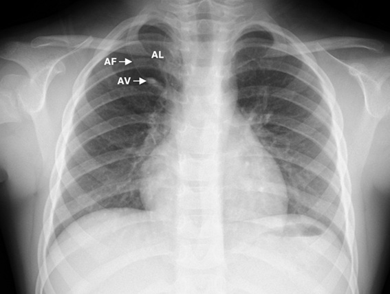
Figure 2. CT scan showing accessory azygos fissure (AF), azygos vein (AV) and azygos lobe (AL).
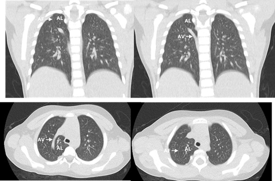
This finding was determined to be a normal anatomical variant, rather than a pathological nodule or mass. No other abnormalities were noted on the CT scan, including no signs of consolidation, pleural effusion, pericardial effusion, or lymphadenopathy. The findings confirmed the benign nature of the lesion and excluded more serious pulmonary conditions.
|
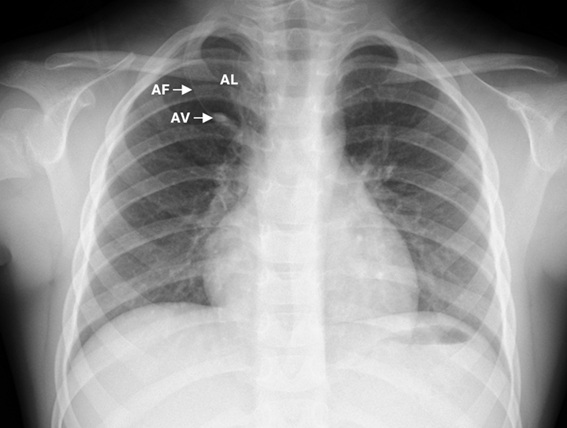
Figure 1. Chest X-ray showing accessory azygos fissure (AF), azygos lobe (AL) and azygos vein (AV).
|
How can the azygos lobe be identified on a chest X-ray or CT scan?
The azygos lobe is not a true lobe of the lungs but rather a rare congenital variant of the lung, typically on the right side, as seen in our case. It is more frequent in males and may have a genetic predisposition.1
Recognizing this anomaly is vital, as it may mimic several pathological conditions, including bullae, abscesses, or lung masses.
The azygos lobe was initially described by the German anatomist Heinrich Wrisberg (1739-1808). It develops when the posterior cardinal vein, which eventually becomes the upper thoracic portion of the azygos vein, fails to migrate properly during fetal development. Typically, this vein moves to its definitive location in the mediastinum, traversing the apex of the right lung. The incidence of this variation is about 1% on CXR and 1.2% on CT scans.2 Chest X-ray is usually the primary diagnostic tool for identifying the azygos lobe.3 As in our case, its radiographic appearance is characterized by a convex line due to the azygos fissure, a triangular area (the trigone) formed by extrapleural tissue along the fissure, and a teardrop shape that connects to the azygos vein in the lower part of the lung.4
The azygos lobe is categorized into three types based on the position of the azygos fissure in relation to the lung apex. Type A is characterized by the trigone being lateral to the apex of the lung; Type B occurs when the trigone is more centrally located, with a more vertical fissure; and Type C is when the trigone is positioned medially, with a fissure extending from the mediastinum.4
While the azygos lobe is typically asymptomatic, it can occasionally be associated with conditions such as malignancies, hemothorax, pneumothorax, vascular anomalies, and even situs inversus totalis. Anatomy of azygos lobe is clinically important during thoracic surgical procedures.3,4 Therefore, it is important to consider the azygos lobe when interpreting posteroanterior CXR which are commonly performed in pediatric clinics. In this case we found azygos lobe incidentally.
In summary, this case underscores the significance of identifying the azygos lobe on chest X-rays and in this way perhaps dispense the CT scan in a healthy and asymptomatic child. It serves as a reminder that this anatomical variation should be included in the differential diagnosis of lung conditions. Notably, treatment is only necessary if an associated pathology is present. |
| |
| Compliance with ethical standards |
|
Funding: None
|
|
|
Conflict of Interest: None
|
|
- Aziz, A., Ashizawa, K., Nagaoki, K., et al. (2004). High resolution CT anatomy of the pulmonary fissures. Journal of Thoracic Imaging, 19(3), 186-191. https://doi.org/10.1097/01.rti.0000125827.01492.34. [CrossRef] [PubMed]
- Ndiaye, A., Ndiaye, N. B., Ndiaye, A., et al. (2012). The azygos lobe: An unusual anatomical observation with pathological and surgical implications. Anatomical Science International, 87(3), 174-178. https://doi.org/10.1007/s12565-012-0144-3. [CrossRef] [PubMed]
- Kobayashi, T., Satoh, K., Kawase, Y., et al. (1995). Bronchial and vascular supply in azygos lobe of inflated fixed lung: A case study. Radiation Medicine, 13, 31-33. https://doi.org/10.1007/BF02999363. [CrossRef]
- Ariyürek, O. M., Gülsün, M., & Demirkazik, F. B. (2001). Accessory fissures of the lung: Evaluation by high-resolution computed tomography. European Radiology, 11, 2449-2453. https://doi.org/10.1007/s003300100838. [CrossRef] [PubMed]
|
|
DOI: https://doi.org/10.7199/ped.oncall.2026.73 |
| |
Cite this article as:
Silva A S d S L d, Malato M T, Ramos D F. Azygos Accessory Fissure in a Pediatric Patient. Pediatr Oncall J. 2025 Mar 11. doi: 10.7199/ped.oncall.2026.73
|