Ashwin Borade1, Ritesh Radhakrishnan Palliana2, D K Singh2.
1Consultant Pediatrician, Inamdar Multispeciality Hospital, Pune,
2Department of Pediatrics, Dr. R.N Cooper Municipal General Hospital, Mumbai, India.
ADDRESS FOR CORRESPONDENCE
Dr. Ashwin Borade , Inamdar Multispeciality Hospital, S.No.15, Fatima Nagar, Pune 411040. Maharashtra, India.
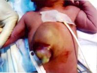
Email: ashwinborade@yahoo.com | | Keywords | | Cantrell's pentalogy, sternal defect. | | | A term male baby weighing 2.5 kg was born to a primi, non-consanguineous married 20-year-old woman. The baby was transferred to our hospital at 18 hrs of age from a peripheral health center. Pregnancy and delivery were supervised and were uneventful. There was no history of a prenatal ultrasound diagnosis of the anomaly. There was no history suggestive of exposure to teratogens during pregnancy. There was no family history of birth defects. The child cried spontaneously at birth. The chest examination revealed an obvious anterior chest wall defect covered by skin, which was pink and membranous over the epigastrium (Figure 1). The cardiac impulse was visible. Palpation of the chest wall revealed a complete sternal defect. Breath sounds were well heard all over the chest, only the first and second heart sounds were heard over the precordium. The heart rate was 140 per minute. The umbilical stump was intact with no obvious anomaly of the umbilicus. The baby also had diaphragmatic hernia. No other anomalies were seen on examination. Echocardiography revealed Tetralogy of Fallot. The child remained in-patient for one week, and 70% alcohol (ethanol) dressing of the membranous covering over the epigastrium was done daily to encourage epithelialization. The child remained clinically stable throughout the period of admission. Child was referred for cardiac surgery at higher center. Patient was lost to follow up.
Fig.1 Anterior chest wall defect
Cantrell's pentalogy is a rare congenital anomaly in which defects of the anterior abdominal wall, sternum, and diaphragm are associated with cardiac anomalies .The reported incidence of CP is 1:100000 births in developed countries.
Cantrell described in 1958 a syndrome in which a ventral diaphragmatic hernia occurred in association with omphalocele. (1) This rare syndrome is now called the pentalogy of Cantrell which consists of the five anomalies includes a deficiency of the anterior diaphragm, a midline supraumbilical abdominal wall defect, a defect in the diaphragmatic pericardium, various congenital intracardiac abnormalities and a defect of the lower sternum. (2) Cantrell's Pentalogy is 2.7 times more common in boys, and African Americans may be more predisposed. (3)
Based on a review of 61 cases of Cantrell's pentalogy, Toyama suggested the following classification for the syndrome: Class 1, certain diagnosis, with all five defects present; Class 2, probable diagnosis, with four defects (including intracardiac and ventral abdominal wall abnormalities) present; and Class 3, incomplete, with various combinations of defects present (but always including a sternal abnormality).(4)
The etiology of the pentalogy is not well established. Cantrell proposed a theory stated that developmental failure of the mesoderm in early embryonic life between 14 and 18 days of gestation results in a failure in the development of the transverse septum of the diaphragm, and of the ventromedial migration of the paired mesodermal folds of the upper abdomen. (1,5) Congenital defects of the sternum may vary from simple notching of the manubrium and irregularities in the shape of the xiphoid to absence of the entire sternum. Abdominal wall defects include omphalocele, diastasis recti, epigastric hernia, umbilical hernia, and combined defects. The most common abdominal wall defect is omphalocele. Deficiencies of the diaphragmatic pericardium and the anterior diaphragm are common defects.
Cardiac lesions may vary widely. Cantrell et al stated that congenital intracardiac anomalies are consistent elements of the pentalogy, with ventricular septal defect in every case (100%), atrial septal defect in 53%, pulmonary stenosis in 33%, tetralogy of Fallot in 20% and left ventricular diverticulum in 20%.
Differential diagnosis includes isolated ectopia cordis, isolated abdominal wall defect, amniotic band syndrome, and body stalk anomaly. The syndrome should be considered with any diagnosis of omphalocele or ectopia cordis. The key features for distinguishing these conditions is the position of abdominal wall defect in relation to the umbilical cord, eviscerated organs, the presence or absence of membranes or bands, and associated anomalies.
The prognosis depends on the severity of the anomalies. Complications include rupture of viscera during delivery, sepsis, cyanosis, congestive heart failure, cardio-respiratory difficulty and cardiac compression. (6) Despite modern surgical standards, Cantrell's syndrome represents a challenge to the surgeon because of the wide spectrum of anomalies, the severity of the abdominal and cardiac malformations, and the high mortality (7). Management includes a complete workup including karyotype and complete ultrasonographic search for other anomalies performed. A multidisciplinary team approach should be employed in the management of these patients. Corrective cardiac surgery would be needed in patients with cardiac anomalies. Successful corrective or palliative cardiothoracic surgery has been reported in these patients. Patients should be reviewed by a pediatric anesthetist before surgery, due to the peculiarities of anesthesia that may arise from the component anomalies. Sowande et al intended a repair of the sternal cleft in these patients, to provide a bony protection for the mediastinal structures (8).
Because of the poor prognosis, early antenatal sonographic detection of it is important and allows for elective abortion before viability. After viability, a periodic ultrasonographic evaluation of the lesions, fetal growth and delivery in a tertiary center is recommended.
Contributors: RP, DKS were involved in management of patient. RP, AB were involved in preparing the manuscript. AB will act as guarantor of the study. | | | | Compliance with Ethical Standards | | Funding None | | | | Conflict of Interest None | | |
- Cantrell JR, Haller JA, Ravitch MM. A syndrome of congenital defect involving the abdominal wall, sternum, diaphragm and heart. Surg Gynecol Obstet. 1958; 107: 602-614. [PubMed]
- Dane C, Dane B, Yayla M, Cetin A. Prenatal diagnosis of a case of pentalogy of Cantrell with spina bifida. J Postgrad Med. 2007; 53: 146-148. [CrossRef]
- Bittmann S, Ulus H, Springer A. Combined pentalogy of cantrell with tetralogy of fallot, and gallbladder agenesis, and polysplenia: a case report. J Pediatr Surg. 2004; 39: 107-109. [CrossRef]
- Toyama WM. Combined congenital defects of the anterior abdominal wall, sternum, diaphragm, pericardium and heart: A case report and review of the syndrome. Pediatrics. 1972; 50: 778-792. [PubMed]
- Haynor DR, Shuman WP, Brewer DK, Mack LA. Imaging of fetal ectopia cordis: roles of sonography and computed tomography. J Ultrasound Med. 1984; 3: 25-27. [CrossRef]
- Marwah VV, Narula MK, Anand R, Solanki R. Pentology of cantrell. Indian J Radiol Imaging. 2002; 12: 450-451.
- Vazquez-Jimenez JF, Muehler EG, Daebritz S, Keutel J, Nishigaki K, Huegel W et al. Cantrell's syndrome: a challenge to the surgeon. Ann Thorac Surg. 1998; 65: 1178-1185. [CrossRef]
- Sowande OA, Anyanwu L, Talabi AO, Babalola OR, Adejuyigbe O. Pentalogy of cantrell: A report of three cases. J Surg Tech Case Report 2010;2:20-3. [CrossRef]
|
| Cite this article as: | | Borade A, Palliana R R, Singh D K. Pentalogy of Cantrell. Pediatr Oncall J. 2011;8: 51-52. |
|