Isabel Ribeiro1, Rita Gomes1, Catarina Mendes Pinto2, Ana Sofia Marinho3, Margarida Paiva Coelho1, Paula Garcia1
1Pediatrics Department, Centro Materno-Infantil do Norte Albino Aroso, Centro Hospitalar Universitário do Porto (CHUPorto), Portugal, 2Department of Neuroradiology, Centro Hospitalar Universitário do Porto (CHUPorto), Portugal, 3Department of Pediatric Surgery, Centro Hospitalar Universitário do Porto (CHUPorto), Portugal
Address for Correspondence: Isabel Ribeiro, Rua Doutor Raúl Chagas, number 109, 4435-124 Porto, Portugal.
Email: isabelm.morais.ribeiro@gmail.com
|
Discussion :
The newborn was admitted to the ward and started on empiric intravenous antibiotic therapy for parotiditis. Due to the absence of clinical improvement at 72 hours, a new ultrasound was performed which had similar findings than the previous one. A magnetic resonance imaging (MRI), performed later, revealed a highly vascularized lobulated lesion, with early uptake in the dynamic study and replacement of the entire glandular stroma, suggestive of a parotid infantile hemangioma (Figure 2). Antibiotic therapy was discontinued and therapy with propranolol was started due to the functional and aesthetic risk of the lesion. Regular ambulatory follow-up was ensured by a multidisciplinary team encompassing pediatricians, hematology and pediatric surgeons. The patient is now in the 4th month of treatment, presenting with substantial involution of the lesion. To this date, still has neutropenia (but no other cytopenias), whose cause hasn´t been identified. There are no infectious complications, growth parameters are appropriate for his age.
Figure 2. MRI A) axial T2 and B) axial T1 with intravenous contrast showing an enhanced lobulated soft tissue mass at the parotid region, suggestive of parotid hemangioma. 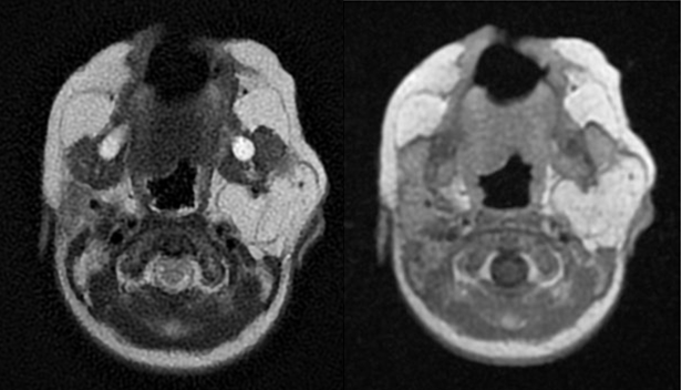
Despite being rare, parotid gland hemangioma is the most common non-inflammatory salivary gland mass in the pediatric population. In this case, the early presentation and the absence of a superficial component made the diagnosis more challenging. Infantile hemangiomas can subside without treatment, but rapid growth may cause aesthetic complications, obstruction and distortion of adjacent structures, as well as lesion ulceration. Clinical suspicion and early diagnosis avoids unnecessary investigation and treatments, enabling the correct therapeutic approach and guidance. | References : | - Abidi KT, Kamal NM, Althobaiti KA, Althobaiti SD, Halabi YA, Alalyani SA. Infantile Parotid Hemangioma With Diagnostic Dilemma: A Case Report. Clin Med Insights Case Rep. 2022 Jan 19;15:11795476211073385. doi: 10.1177/11795476211073385. PMID: 35095285; PMCID: PMC8793416.
- Cheng CE, Friedlander SF. Infantile hemangiomas, complications and treatments. Semin Cutan Med Surg. 2016 Sep;35(3):108-16. doi: 10.12788/j.sder.2016.050. PMID: 27607318.
- Mantadakis E, Tsouvala E, Deftereos S, Danielides V, Chatzimichael A. Involution of a large parotid hemangioma with oral propranolol: an illustrative report and review of the literature. Case Rep Pediatr. 2012;2012:353812. doi: 10.1155/2012/353812. Epub 2012 Nov 25. PMID: 23227404; PMCID: PMC3512253.
- Munden A, Butschek R, Tom WL, Marshall JS, Poeltler DM, Krohne SE, Alió AB, Ritter M, Friedlander DF, Catanzarite V, Mendoza A, Smith L, Friedlander M, Friedlander SF. Prospective study of infantile haemangiomas: incidence, clinical characteristics and association with placental anomalies. Br J Dermatol. 2014 Apr;170(4):907-13. doi: 10.1111/bjd.12804. PMID: 24641194; PMCID: PMC4410180.
|
|
| Correct Answers : |  100% 100% |
Last Shown : Jul 2025
|