Lamis Al Sawafi1, Nasser Al Shafouri1, Mahmoud Khalid1, Farida AmbuSaidi2.
1Department of Pediatrics, Ibri Hospital, Ministry of Health, Ibri, Oman,
2Department of Radiology, Ibri Hospital, Ministry of Health, Ibri, Oman.
ADDRESS FOR CORRESPONDENCE
Nasser Al Shafouri, Department of Pediatrics, Ibri Hospital, Ministry of Health, PO Box 618, Postal Code 511, Ibri, Sultanate of Oman.
Email: alshafouri@gmail.com | | Abstract | | Neonatal arterial thrombosis is rare, with an estimated incidence of 0.5 per 10,000 live births, and is usually iatrogenic; spontaneous thrombosis occurring immediately at birth is exceptionally uncommon. We report a male infant born at 37 weeks’ gestation by emergency caesarean section for placental abruption. At six minutes of life, examination revealed marked pallor of the left lower limb with absent left femoral and pedal pulses, indicating acute limb ischaemia. Doppler ultrasound confirmed complete occlusion of the left external iliac artery by a 3-cm thrombus with absent distal flow. The mother had diet-controlled gestational diabetes mellitus, sickle cell trait, and a previous pregnancy complicated by abruption and pre-eclampsia with HELLP syndrome. After exclusion of intracranial haemorrhage and multidisciplinary consultation, continuous intravenous unfractionated heparin was commenced and titrated to a therapeutic activated partial thromboplastin time. Limb perfusion improved within four hours, the femoral pulse became palpable by 24 hours, and serial Doppler studies showed complete recanalisation within seven days without bleeding complications. The infant was discharged on day seven with normal limb perfusion, and subsequent thrombophilia screening was normal. This case highlights that spontaneous neonatal external iliac artery thrombosis, although rare, requires prompt clinical recognition and urgent Doppler evaluation, and that early anticoagulation with unfractionated heparin can achieve complete thrombus resolution and limb preservation without surgery or thrombolysis. | | | | Keywords | | Neonatal thrombosis, External iliac artery, Anticoagulation, heparin, Limb ischaemia. | | | | Introduction | | Neonatal arterial thrombosis is an uncommon but potentially limb- and life-threatening condition, with an estimated incidence of 0.5 per 10,000 live births.1 The neonatal period represents a prothrombotic state characterised by hemostatic immaturity, relative deficiency of natural anticoagulants, and physiological elevation of procoagulant factors.2 Iatrogenic factors, particularly umbilical arterial catheterisation and central venous access, account for approximately 90% of neonatal arterial thrombosis cases.3 Spontaneous thrombosis occurring immediately at birth, without any preceding vascular intervention, is very rare. Additional risk factors include perinatal asphyxia, sepsis, polycythaemia, congenital heart disease, and maternal diabetes mellitus4, while inherited thrombophilias further increase risk.5 Doppler ultrasonography is the diagnostic modality of choice, and anticoagulation with unfractionated or low-molecular-weight heparin remains the mainstay of therapy; surgery and thrombolysis are reserved for refractory, limb-threatening cases.1,3 We describe a rare case of spontaneous neonatal external iliac artery thrombosis managed successfully with early anticoagulation. | | | | Case Report | A male infant was delivered at 37 weeks’ gestation by emergency lower-segment caesarean section for placental abruption. Birth weight was 3.02 kg, length 50 cm, and head circumference 34 cm. Apgar scores were 8 and 9 at one and five minutes, respectively. The mother was a 31-year-old, gravida 2 para 1, with diet-controlled gestational diabetes mellitus and sickle cell trait; her previous pregnancy had been complicated by placental abruption and pre-eclampsia with HELLP syndrome.
At six minutes of age, examination revealed marked pallor of the left lower extremity extending to the lower abdomen (Figure 1), and the infant was transferred to the neonatal intensive care unit. He was well-appearing with mild tachypnoea and oxygen saturation of 96% in room air. The left femoral and pedal pulses were absent, with a cold, pale left lower limb and capillary refill exceeding five seconds; the right lower limb was normally perfused. A grade 2/6 systolic murmur was noted, and neurological examination was normal.
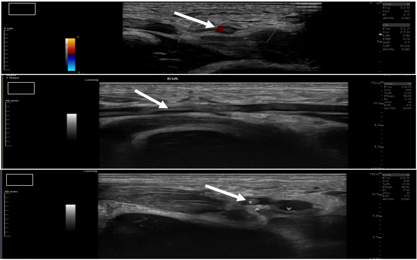
Doppler ultrasound at one hour of life demonstrated complete occlusion of the left external iliac artery by a 3-cm hyperechoic thrombus with absent distal flow; the left femoral and popliteal arteries were patent but showed minimal flow signal (Figure 2). Investigations showed haemoglobin 19.4 g/dL, haematocrit 61.7%, platelets 228 × 10⁹/L, prothrombin time 12 s, activated partial thromboplastin time 35 s, and fibrinogen 2.1 g/L; arterial lactate was 4.0 mmol/L. Echocardiography showed a 6-mm secundum atrial septal defect, a small patent ductus arteriosus, and normal ventricular function without intracardiac thrombus. Cranial ultrasound was normal.
Figure 1. Clinical photograph taken at six minutes of life showing marked pallor of the left lower extremity extending to the lower abdomen, consistent with acute left lower limb ischaemia secondary to external iliac artery thrombosis. A black bar has been placed over the genitalia; the infant is otherwise non-identifiable.
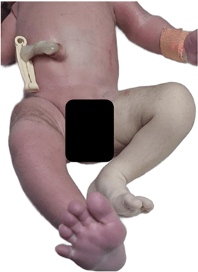
Figure 2. Doppler ultrasound images demonstrating thrombosis of the left external iliac artery (EIA); white arrows indicate the hyperechoic thrombus. (A) Axial view showing marginal flow within the affected artery. (B) Coronal view demonstrating an approximately 3-cm-long thrombus in the left EIA. (C) Axial view confirming intraluminal thrombus.
After multidisciplinary consultation and exclusion of intracranial haemorrhage, continuous intravenous unfractionated heparin was commenced at 28 units/kg/hour and titrated to a therapeutic activated partial thromboplastin time (Table 1). Limb colour and temperature improved within four hours, and the left femoral pulse became palpable with normal capillary refill by 24 hours. Serial Doppler studies showed progressive thrombus resolution, with complete recanalisation by day seven and no bleeding complications. Respiratory support was weaned by day three and full enteral feeding established by day five. heparin was continued for seven days, and the infant was discharged on day seven with patent aorta, iliac, femoral, and popliteal arteries on Doppler ultrasound. Subsequent thrombophilia screening was normal, and the child remained well at follow-up with no recurrence of limb discolouration.
Table 1. Unfractionated heparin dose adjustment protocol based on the activated partial thromboplastin time (aPTT).
| aPTT (seconds) |
aPTT ratio |
Dose adjustment |
Repeat aPTT |
| <50 |
<1.5 |
Bolus 50 units/kg; increase infusion by 10% |
4 hours |
| 50–59 |
1.5–1.9 |
Increase infusion by 10% |
4 hours |
| 60–85 |
2.0–2.7 |
No change (therapeutic range) |
24 hours |
| 86–95 |
2.8–3.0 |
Decrease infusion by 10% |
4 hours |
| 96–120 |
3.0–3.8 |
Hold infusion 30 min; decrease by 10% |
4 hours |
| >120 |
>3.8 |
Hold infusion 60 min; decrease by 15% |
4 hours |
aPTT: activated partial thromboplastin time. Therapeutic aPTT range 60–85 seconds (ratio 2.0–2.7). Initial maintenance dose 28 units/kg/hour. Repeat aPTT four hours after each dose adjustment.
| | | | Discussion | This case represents an exceptionally rare presentation of spontaneous neonatal external iliac artery thrombosis occurring immediately at birth. Several recognised risk factors were present. Maternal gestational diabetes increases neonatal thrombotic risk through endothelial dysfunction, increased platelet aggregation, and fetal polycythaemia6, and the infant’s high haematocrit (61.7%) is consistent with this mechanism. Placental abruption may contribute through release of tissue factor and activation of the coagulation cascade7, and the mildly elevated lactate suggests a degree of perinatal hypoxia that can further promote endothelial activation.
Doppler ultrasound remains the diagnostic modality of choice for suspected neonatal limb ischaemia, offering high sensitivity without radiation exposure.1 The onset of signs within minutes of birth underscores the importance of immediate clinical assessment and imaging in any neonate with perfusion abnormalities. Unfractionated heparin is generally preferred as first-line therapy because of its rapid onset, reversibility, and extensive monitoring experience in neonates3, and the prompt response in our patient supports early anticoagulation for limb preservation. Thrombolysis and surgery are reserved for threatened limb viability or failure of anticoagulation8, neither of which was required here.
Prognosis depends on the extent of initial ischaemia, timeliness of intervention, and any underlying thrombophilia5,9; the complete resolution and normal screening in this case indicate an excellent outlook. Our report adds to the limited literature on spontaneous neonatal arterial thrombosis: Tsonis et al. described a comparable spontaneous femoral artery thrombosis successfully treated with anticoagulation10, and Makwana et al. similarly reported reversal of ischaemia without long-term sequelae.8 | | | | Conclusion | | Spontaneous neonatal external iliac artery thrombosis is extremely rare but serious, requiring immediate recognition and treatment. Early anticoagulation with unfractionated heparin can achieve complete thrombus resolution and limb preservation without surgery or thrombolysis. Clinicians should maintain a high index of suspicion for arterial thrombosis in any neonate with acute limb ischaemia, particularly in the presence of maternal diabetes, placental complications, or a family history of thrombosis. | | | | Compliance with Ethical Standards | | Funding None | | | | Conflict of Interest None | | |
- Aslam M, Guglietti D, Hansen AR. Neonatal arterial thrombosis at birth: case report and literature review. Am J Perinatol 2008;25:347-52. [CrossRef] [PubMed]
- Nowak-Göttl U, Janssen V, Manner D, Kenet G. Venous thromboembolism in neonates and children - update 2013. Thromb Res 2013;131 Suppl 1:S39-41. [CrossRef] [PubMed]
- Rashish G, Paes BA, Nagel K, et al. Spontaneous neonatal arterial thromboembolism: infants at risk, diagnosis, treatment, and outcomes. Blood Coagul Fibrinolysis 2013;24:787-97. [CrossRef] [PubMed]
- Tolbert J, Carpenter SL. Common acquired causes of thrombosis in children. Curr Probl Pediatr Adolesc Health Care 2013;43:169-77. [CrossRef] [PubMed]
- Kenet G, Aronis S, Berkun Y, et al. Impact of inherited thrombophilia on arterial ischemic stroke and cerebral sinovenous thrombosis in children: a systematic review and meta-analysis. Circulation 2010;121:1838-47. [CrossRef] [PubMed] [PMC free article]
- Mitanchez D, Yzydorczyk C, Simeoni U. What neonatal complications should the pediatrician be aware of in case of maternal gestational diabetes? World J Diabetes 2015;6:734-43. [CrossRef] [PubMed] [PMC free article]
- Lykke JA, Bare LA, OlsenJ, et al. Thrombophilias and adverse pregnancy outcomes: results from the Danish National Birth Cohort. J Thromb Haemost 2012;10:1320-5. [CrossRef] [PubMed]
- Makwana BM, Rabadia PJ. Neonatal arterial thrombosis at birth. Int J Contemp Pediatr 2023;10:1469-71. [CrossRef]
- Ichiyama M, Ohga S, Ochiai M, et al. Age-specific onset and distribution of natural anticoagulant deficiency in pediatric thromboembolism. Pediatr Res 2016;79:81-6. [CrossRef] [PubMed]
- Tsonis O, Gouvias T, Gkrozou F, et al. Neonatal femoral artery thrombosis at the time of birth: a case report. J Pediatr Neonat Individ Med 2020;9:e090214.
DOI: https://doi.org/10.7199/ped.oncall.2027.18
|
| Cite this article as: | | Sawafi L A, Shafouri N A, Khalid M, AmbuSaidi F. Spontaneous External Iliac Artery Thrombosis in a Newborn: Successful Management with Early Anticoagulation. Pediatr Oncall J. 2026 Jul 21. doi: 10.7199/ped.oncall.2027.18 |
|