R Nivetha, T Palanivelraja, A Srenivas, S Gobinathan, K.S Kumaravel.
Department of Pediatrics, Govt. Mohan Kumaramangalam Medical College, Salem, India.
ADDRESS FOR CORRESPONDENCE
K. S. Kumaravel, 191A, Shankar Nagar, Salem, Tamil Nadu, India. PIN: 636 007.
Email: kumaravelks@rediffmail.com | | Abstract | Introduction: The Umbilical Vein Catheter (UVC) tip should ideally be seen radiologically at the level of the diaphragm or the T9 vertebra, and an incorrect position is associated with complications. This study aims to estimate the accuracy and safety of Real-Time ultrasound guided Umbilical Venous Catheterisation (RTUVC) in correctly placing UVC when compared to the conventional landmark method.
Methods: This is a pre-post study conducted in an urban hospital before and after the implementation of RTUVC. Neonates who had UVC inserted before the implementation were taken as controls, and neonates who had UVC inserted after the implementation of RTUVC were taken as cases. The primary outcome measure was the correct positioning of UVC in the first attempt. The secondary outcome measures were the number of X-rays obtained, the need for repositioning, the number of UVC withdrawals, and portal vein entry.
Results: There was no statistically significant difference between the groups in gender, gestational age, birth weight, or mode of delivery. The correct placement of UVC was achieved in a significantly higher number of neonates in the RTUVC group than the controls (90% vs 66.7%) (P=0.006). The UVC repositioning times and mean number of X-rays taken were lower in the RTUVC group. There were no UVC withdrawals and portal vein entry observed in the RTUVC group.
Conclusion: This study has demonstrated that the implementation of RTUVC significantly improved the correct placement of UVC on the first attempt and resulted in no complications related to portal vein entry and catheter withdrawals. | | | | Keywords | Portal Vein, Inferior Vena Cava, Shukla’s formula, Dunn formula, Ultrasound.
| | | | Introduction | Securing vascular access is an important step in the management of critically ill and preterm neonates.1 Umbilical Venous Catheter (UVC) is commonly used for venous access in most neonatal intensive care units (NICU). It can be used to administer fluids, blood, drugs, and parenteral nutrition. The UVC insertions are done routinely in NICUs by the residents and neonatologists after formal training. Various formulas are available for determining the length of the UVC to be inserted.2,3,4 The Shukla’s formula is based on birth weight, and the Dunn’s formula is based on the shoulder-umbilical length. A study by Lean et al demonstrated that none of these formulas is adequate for the correct positioning of UVC, and it needs some repositioning to place it correctly.5
The UVC tip should ideally be placed at the junction of the Right Atrium (RA) and Inferior Vena Cava (IVC).6 There are some conventional ways to confirm the UVC tip position radiologically, using the diaphragm or the vertebral bodies as a landmark. The UVC tip should ideally be seen at the level of the diaphragm or at the T9 vertebra.7 The incorrect position of the UVC was associated with various complications.1 The placement of UVC higher up at the right atrium was associated with myocarditis, arrhythmias and tamponade.8,9 On the other hand, the placement of UVC at a lower level was associated with portal vein thrombosis, portal hypertension and hepatic necrosis.10 Hence, it is important to ensure that the UVC is correctly placed.
With the increasing use of Point of Care Ultrasound (POCUS) in NICUs, confirming the UVC tip can now also be done with ultrasound.1 However, both radiological and ultrasound confirmation require repositioning or withdrawal if the UVC is placed incorrectly.1 To address these limitations, researchers proposed Real-Time ultrasound-guided UVC (RTUVC) insertion.11 In RTUVC, the UVC insertion is performed under ultrasound guidance—one operator inserts the catheter while another visualizes its position in real time. Adjustments are made during the procedure, eliminating the need for radiological confirmation. The RTUVC reduces turnaround time and radiation exposure compared to the conventional landmark method for UVC confirmation. Although many studies have evaluated RTUVC's effectiveness, it has not yet become standard practice in many NICUs.12 The success of RTUVC largely depends on the expertise of the sonographer and the clinician performing the insertion, as both must coordinate effectively. Limited data are available in India regarding the superiority of RTUVC over the traditional landmark method.
Aims and objectives
This study aims to estimate the accuracy and safety of RTUVC in correctly placing UVC when compared to the conventional landmark method of confirmation.
| | | | Methods | This is a pre-post study conducted in the NICU of an urban hospital before and after the implementation of RTUVC. The study hospital implemented RTUVC as a standard protocol in January 2026. Neonates who had UVC inserted before the implementation of RTUVC from September 2025 to December 2025 were included as controls (historical controls). Neonates who had UVC inserted after the implementation of RTUVC from January 2026 to April 2026 were included as cases. Neonates with shock, major congenital malformations, and congenital heart diseases were excluded from both groups. The Institutional Human Ethics Committee approved the study. Consent was obtained from the parents.
The RTUVC procedure was performed by 2 trained faculty members – one operator (2nd Author) who inserted the UVC, and another sonographer (4th Author) who visualised the umbilical vein and guided the operator to ensure correct placement. The sonogram was performed by a pediatrician with experience in POCUS and the operator who inserted the UVC is an experienced pediatrician. For all the neonates included in the study, RTUVC was done by the same operator and sonographer to avoid inter-operator variability. The RTUVCs done by other operators/sonographers were excluded.
Figure 1. Flow chart of study participants.
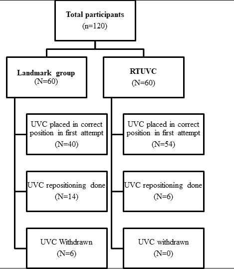
RTUVC: Real-Time ultrasound guided Umbilical Vein Catheterisation,
UVC: Umbilical Vein Catheter
Controls
The data for the controls were obtained from the case records, which included name, gestational age, birth weight, morbidity, radiologic umbilical tip position (correct, high, or low placement), number of X-rays taken, number of repositioning attempts, withdrawal, and portal vein entry. In the unit, UVCs were inserted under aseptic precautions, using Modified Shukla’s formula ((3 × weight in kg + 9) / 2 cm) to determine insertion length.5 The size of the UVC was based on the neonate’s weight. Radiologic confirmation of the UVC tip position was performed immediately. If placement was incorrect, repositioning was performed, and radiologic confirmation was repeated. For neonates in whom portal vein entry of the UVC was observed, the UVC was removed, and vascular access was established via peripheral veins as per the unit’s protocol.
Cases
The RTUVC insertion was performed using the Mindray MX7 ultrasound system with a P10-4s neonatal cardiac/sector probe operating at a frequency range of 5-10 MHz. The subxiphoid (subcostal) window was used for real-time visualisation and guidance during catheter advancement. Ultrasound examination enabled identification of the umbilical vein, left portal vein, ductus venosus, inferior vena cava (IVC), right atrium (RA), and surrounding liver parenchyma during the procedure. Real-time imaging was utilised to navigate the catheter course and confirm optimal tip placement at the IVC/RA junction, while also helping to identify any malposition into the portal vein system or intracardiac structures. To further ease the identification of the tip, a saline bolus was used in some cases. In neonates who had RTUVC, radiological confirmation was done, and similar data, as described for controls, were collected prospectively.
Outcome measures
The primary outcome measure was the correct positioning of UVC at the IVC/RA junction in the first attempt. The secondary outcome measures were the number of X-rays obtained, the need for repositioning, the number of UVC withdrawals, and portal vein entry. The difficulties observed during the RTUVC insertion were also collected.
Sample size calculation
The sample size was calculated based on the primary outcome of correct UVC tip placement at the IVC/RA junction. Based on studies by Luo et al., the success rate of the conventional landmark method was estimated at 68%, whereas a study by Rubortone et al. reported that the real-time ultrasound-guided (RTUVC) method was expected to achieve 90% success.13,14 To detect this 22% difference, a minimum of 53 neonates per group was required with 80% power at a two-sided alpha of 0.05, using the standard formula for comparison of two independent proportions:
n = [Zα/2·√(2p̄(1-p̄)) + Zβ·√(p₁(1-p₁)+p₂(1-p₂))]² / (p₁-p₂)²
To allow for potential exclusions and to improve precision, 60 neonates per group were enrolled, providing approximately 85% power for this comparison.
Statistical analysis
For statistical analysis, the R software was used. Baseline categorical variables were compared using the Chi-square test. Continuous baseline variables (gestational age and birth weight) were non-normally distributed on the Shapiro-Wilk test (p <0.05 for all), so the non-parametric Mann-Whitney U test was used for between-group comparison, and results are reported as mean ± SD for descriptive purposes. The primary outcome and categorical secondary outcomes (need for repositioning, portal vein entry, and catheter withdrawal) were analysed using Fisher’s exact test, and the numeric count of confirmation X-rays required per neonate was treated as an ordinal count variable and compared between the groups using the two-tailed Mann-Whitney U test. Statistical significance was defined as a P-value ≤0.05 in all analyses. | | | | Results | Table 1. Baseline characteristics of the study population.
| Variable |
Landmark method
(n=60) |
RTUVC method
(n=60) |
P Value |
| Mean Gestational age (weeks) |
31.4±2.8 |
32.3±3.1 |
0.061$ |
| Mean Birth weight (g) |
1490.0±534.4 |
1519.8±595.9 |
0.831$ |
| Gender |
| Male |
28 |
32 |
0.583* |
| Female |
32 |
28 |
|
| Mode of delivery |
| Labour Natural |
26 |
28 |
0.854* |
| LSCS |
34 |
32 |
|
$Mann-Whitney U test, *Chi-square test
The baseline data are tabulated in Table 1. Among the neonates who underwent RTUVC insertions during the study period, 60 neonates were enrolled. Sixty neonates were identified retrospectively as historical controls between September 2025 and December 2025. The mean birth weights were 1490.0 ± 534.4 and 1519.8 ± 595.9 for the controls and cases, respectively. The mean gestational ages were 31.4 ± 2.8 and 32.3 ± 3.1for controls and cases, respectively. There was no statistically significant difference between the groups in gender, gestational age, birth weight, or mode of delivery.
Table 2. Comparison of outcome measures between the groups.
| Outcome measure |
Landmark method
(n=60) |
RTUVC method
(n=60) |
P Value |
| Correct UVC tip placement on the first attempt |
40 (66.7%) |
54 (90%) |
0.006* |
| UVC Repositioning times (%) |
15 (25%) |
6 (10%) |
0.052* |
| UVC withdrawal times (%) |
6 (10%) |
0 |
0.027* |
| Mean Number of X-rays |
1.25 |
1.10 |
0.048$ |
| Portal vein entry (%) |
6 (10%) |
0 |
0.027* |
*Fisher's exact test, $Mann-Whitney U test
Figure 2. Ultrasound image of Umbilical Vein Catheter in the Right Atrium/Inferior Vena Cava Junction.
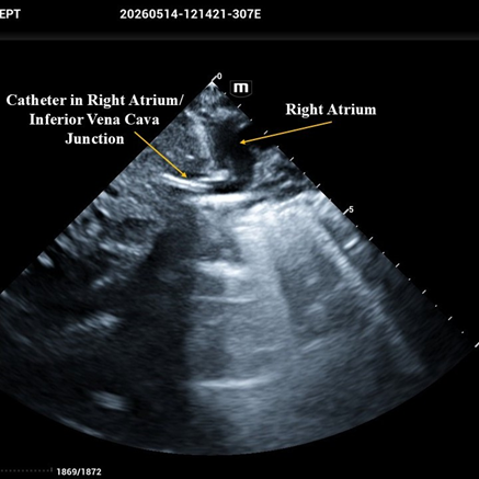
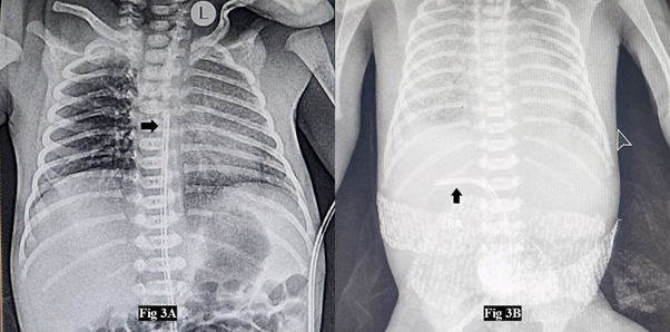
The comparison of outcomes between the groups is tabulated in Table 2. The UVC tip was correctly placed in 66.7% of neonates on the first attempt using the Landmark method, whereas in the RTUVC group, it was placed in 90% of neonates on the first attempt (Figure 2) (P = 0.006). In the landmark group, the UVC tip was found to be low-lying in 13 neonates, and in one neonate, it was found in the right atrium (Figure 3A). In the RTUVC group, difficulty was encountered in detecting the UVC tip in six neonates due to motion artefacts from the crying neonates or bowel gas interfering with the sonographic window. The need for UVC repositioning after failed radiological confirmation was less in the RTUVC group than the landmark group (10% vs 25%), though this did not reach conventional statistical significance (P = 0.052). In all six cases in the RTUVC group, repositioning was done successfully in the second attempt. However, in the landmark group out of 14 cases, in 13 cases, repositioning was done in the second attempt, and in one neonate, correct placement was attained only in the third attempt. There were no UVC withdrawals in the RTUVC group, whereas in 6 neonates, UVC was withdrawn due to a complication of portal vein entry in the Landmark group (Figure 3B). The mean number of X-rays taken was significantly higher in the landmark group (P = 0.048). Complications of portal vein entry of UVC were observed in 6 neonates in the Landmark group, whereas no such cases were reported in the RTUVC group.
Figure 3. Umbilical Vein Catheter Malpositions: 3A: Right Atrium, 3B: Left Portal Vein.
 | | | | Discussion | The use of POCUS in neonatology is expanding every day.15 Also, residents and consultants receive POCUS training upon recruitment to the NICU.16 Many procedures that previously required X-rays were now performed with POCUS. In this study, we evaluated the efficacy and safety of the RTUVC in a single centre. We observed that RTUVC significantly increased the correct placement of UVC on the first attempt, and serious complications such as portal vein entry were absent.
In the present study, the placement of UVC in the correct position in the first attempt was 66.7% with Modified Shukla’s formula. This varies across studies, from 17.8% to 57%.12 The variations may be due to factors like the formula and definition (T8-T10 vertebra, 1 cm above diaphragm, IVC/RA junction) used. The present study improved the correct placement of UVC from 66.7% to 90% in the first attempt. This is consistent with other studies. In a study by Rossi et al. the correct placement improved from 46.2% to 58.8% by using RTUVC.17 In a study by Kaur et al. the accuracy improved from 26% to 56.7% by using RTUVC.18
In this study, we employed RTUVC to help navigate the catheter across the ductus venosus and IVC in real time, which was time-consuming. Though this procedure takes a little more time, the accuracy is higher. In most studies, ultrasound was used to identify the UVC tip at the IVC/RA junction without helping in the navigation of the catheter.17,18 The need for repositioning was lower in the RTUVC group in our study. This was consistent with other studies.12,19 Further, in our study, the mean number of X-rays taken was significantly lower in the RTUVC group. Flaming et al. reported a reduction from 4.1 in the landmark group to 2.3 in the ultrasound group.19 A study by Rossi et al. reported a reduction from 1.5 to 1.19 after employment of ultrasound to guide UVC insertion.17 The reduction in X-rays not only reduces irradiation but also reduces the expenditure.17
In the present study, in the RTUVC group, we encountered difficulty in detecting the UVC by ultrasound in 6 neonates due to the interposition of gastric air and due to the motion artefacts created by the crying neonate. The interference due to the bowel gas was well documented in many studies.11 There are also reports of migration of the UVC after insertion, ranging from 50% to 90%.20,21 In these situations, UVC tip localisation by ultrasonogram will be of great importance, as it can be frequently repeated.1 The future of medical education in UVC insertion depends on the dissemination of knowledge about the RTUVC among all the recruits into the NICU.
The study has a few limitations. This is a single-centre pre-post study with a single operator-sonographer dyad, and hence, the efficacy of RTUVC insertions by other operators/sonographers was not studied. The other variables, like Central Line Associated Blood Stream Infections, turnaround time, and cost involved for X-rays and RTUVC, were not studied. | | | | Conclusion | | In this study, we have demonstrated that the implementation of RTUVC significantly improved the correct placement of UVC in the first attempt. It also resulted in fewer complications, such as portal vein entry and no catheter withdrawals. With adequate training, RTUVC is poised to become a standard of correct UVC placement in all NICUs. | | | | Compliance with Ethical Standards | | Funding None | | | | Conflict of Interest None | | |
- D'Andrea V, Prontera G, Rubortone SA, Pezza L, Pinna G, Barone G, Pittiruti M and Vento G (2022) Umbilical Venous Catheter Update: A Narrative Review Including Ultrasound and Training. Front. Pediatr. 9:774705. doi: 10.3389/fped.2021.774705. [CrossRef] [PubMed] [PMC free article]
- Dunn PM. Localization of the umbilical catheter by post-mortem measurement. Arch Dis Child. (1966) 41:69-75. doi: 10.1136/adc.41.215.69 [CrossRef] [PubMed] [PMC free article]
- Rapid estimation of insertional length of umbilical catheters in newborns. Am J Dis Child. (1986) 140:786-8. doi: 10.1001/archpedi.1986.02140220068034 [CrossRef] [PubMed]
- Verheij GH, te Pas AB, Smits-Wintjens VE, Šràmek A, Walther FJ, Lopriore E. Revised formula to determine the insertion length of umbilical vein catheters. Eur J Pediatr. (2013) 172:1011-5. doi: 10.1007/s00431-013-1981-z. [CrossRef] [PubMed]
- Lean WL, Dawson JA, Davis PG, Theda C, Thio M. Accuracy of five formulae to determine the insertion length of umbilical venous catheters. Arch Dis Child Fetal Neonatal Ed. (2019) 104(2):F165-9. doi: 10.1136/archdischild-2017-314280. [CrossRef] [PubMed]
- R N, R A, S G, K S K. Misdirected Umbilical Venous Catheter in a neonate: A case report and anatomical review. Pediatr Oncall J. 2026 Mar 26. doi: 10.7199/ped.oncall.2026.94 [CrossRef]
- Hoellering AB, Koorts PJ, Cartwright DW, Davies MW. Determination of umbilical venous catheter tip position with radiograph. Pediatr Crit Care Med. (2014) 15:56-61. doi: 10.1097/PCC.0b013e31829f5efa. [CrossRef] [PubMed]
- Sheta A, Al-Awad EH, Soraisham AS. Supraventricular tachycardia associated with umbilical venous catheterization in neonates. J Clin Neonatol. (2018) 7:166-9. doi: 10.4103/jcn.JCN_127_17 [CrossRef]
- Traen M, Schepens E, Laroche S, van Overmeire B. Cardiac tamponade and pericardial effusion due to venous umbilical catheterization. Acta Paediatr. (2005) 94:626-8. doi: 10.1111/j.1651-2227.2005.tb01950.x [CrossRef] [PubMed]
- Chen HJ, Chao HC, Chiang MC, Chu SM. Hepatic extravasation complicated by umbilical venous catheterization in neonates: a 5-year, single-center experience. Pediatr Neonatol. (2020) 61:16-24. doi: 10.1016/j.pedneo.2019.05.004. [CrossRef] [PubMed]
- Cossovel F, Galdo F, Ronfani L, Travan L, Bibalo C and Trappan A (2025) Real-time ultrasound enhances umbilical venous catheters placement in preterm newborns. Front. Pediatr. 13:1567586. doi: 10.3389/fped.2025.1567586. [CrossRef] [PubMed] [PMC free article]
- Ponin L, Ruangkit C, Ruangwattanapaisarn N, Nuntnarumit P. Real-time ultrasound to assess the umbilical catheter position in neonates: a randomized, controlled trial. J Perinatol. 2025 Feb;45(2):235-241. doi: 10.1038/s41372-024-02128-6. Epub 2024 Oct 9. [CrossRef] [PubMed] [PMC free article]
- Luo J, Zheng X, Yang Z, Li K, Chen L, Hei M. Comparative analysis of formulae for umbilical venous catheterization depth based on birth weight. Pediatric Investigation. 2024 Dec 25;8(04):265-70. [CrossRef] [PubMed] [PMC free article]
- Rubortone SA, Costa S, Perri A, D'Andrea V, Vento G, Barone G. Real-time ultrasound for tip location of umbilical venous catheter in neonates: a pre/post intervention study. Ital J Pediatr. 2021 Mar 18;47(1):68. [CrossRef] [PubMed] [PMC free article]
- Stewart DL, Elsayed Y, Fraga MV, Coley BD, Annam A, Milla SS. Use of point-of-care ultrasonography in the NICU for diagnostic and procedural purposes. Pediatrics. 2022;150(6):e2022060053. [CrossRef] [PubMed]
- Kumaravel KS, Anurekha V, Gobinathan S, Palanivelraja T, Aiswarya R, Balaji M. Knowledge Dissemination of Lung Ultrasound in Diagnosing Neonatal Respiratory Distress Syndrome: A Mixed-methods Study. Apollo Medicine. 2026:09760016251413857. [CrossRef]
- Rossi S, Jogeesvaran KH, Matu E, Khan H, Grande E, Meau-Petit V. Point-of-care ultrasound for neonatal central catheter positioning: impact on X-rays and line tip position accuracy. Eur J Pediatr. 2022;181:2097-108. [CrossRef] [PubMed]
- Kaur A, Manerkar S, Patra S, Kalamdani P, Kalathingal T, Mondkar J. Ultrasound-Guided Umbilical Venous Catheter Insertion to Reduce Rate of Catheter Tip Malposition in Neonates: A Randomized, Controlled Trial. Indian J Pediatr.2022;89:1093-98. [CrossRef] [PubMed]
- Fleming SE, Kim JH. Ultrasound-guided umbilical catheter insertion in neonates. J Perinatol. 2011;31:344-49. [CrossRef] [PubMed]
- Dubbink-Verheij GH, Visser R, Tan RNGB, Roest AAW, Lopriore E, TePas AB. Inadvertent migration of umbilical venous catheters often leads to malposition. Neonatology. (2019) 115:205-10. doi: 10.1159/000494369. [CrossRef] [PubMed] [PMC free article]
- Hoellering A, Tshamala D, Davies MW. Study of movement of umbilical venous catheters over time. J Paediatr Child Health. (2018) 54:1329-35. [CrossRef] [PubMed]
DOI: https://doi.org/10.7199/ped.oncall.2028.1
|
| Cite this article as: | | Nivetha R, Palanivelraja T, Srenivas A, Gobinathan S, Kumaravel K. Efficacy and Safety of Real-Time Ultrasound-Guided vs Conventional Landmark Method for Umbilical Venous Catheterisation in Neonates: A Single-Centre Experience. Pediatr Oncall J. 2026 Aug 17. doi: 10.7199/ped.oncall.2028.1 |
|