|
Male Epispadias - Recognition at a glance
Machado Morais Joana1, Martins Filipa1, Coelho Ana2, Mota Céu1, Rocha Sandra1, Pinho Liliana1.
1Neonatal Intensive Care Unit, Centro Materno Infantil do Norte, Porto-Portugal,
2Surgery Department, Centro Materno Infantil do Norte, Porto-Portugal.
ADDRESS FOR CORRESPONDENCE
Joana Machado Morais, Avenida da República, nº81, 4º esquerdo, Matosinhos, 4450-241, Portugal.
Email: machado_joanaf@hotmail.com
Extrophy-Epispadias Complex, Neonatology, Pediatric Surgery
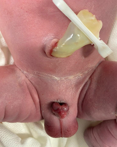
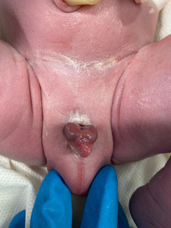
A male newborn delivered at 392/7 weeks to a 28-year-old gravida 1, para 0 woman. Pregnancy was uneventful and no fetal anomalies were detected at prenatal ultrasound. The mother underwent a cesarean section due to pelvic presentation. Apgar score was 9, 10, 10 at 1, 5 and 10 minutes, respectively. Anthropometric measurement at birth was appropriate for gestational age (weight 3090 g, head circumference 34.5 cm, length 46 cm). The postnatal exam showed a dorsally open urethral meatus with mild pubic diastasis and closed anterior abdominal wall and bladder, compatible with epispadias diagnosis. The neonate exhibited micrognathia, with no other significant abnormalities. Renal and cranial ultrasounds were normal. Genetic tests revealed a normal karyotype, but an abnormal array result (13q34x3, 16p13.3x1). During the first year, the patient will remain under surveillance, assessing bladder capacity by voiding cystourethrography and cystoscopy, whose findings will help deciding the type of surgery. The neonate will need surgical repair of epispadias and, possibly, bladder neck repair.
Figure 1. Male Epispadias.

Figure 2. Male Epispadias.
|
Figure 1. Male Epispadias
|
What is the diagnosis?
The Extrophy-Epispadias Complex (EEC) is a spectrum of rare abdominal midline malformations, affecting the urinary system, musculoskeletal system, pelvis, pelvic floor, abdominal wall, genitalia and sometimes the spine and the anus.1,2 It comprises different levels of severity, from epispadias, the mildest form, classical bladder exstrophy to exstrophy of the cloaca, the most severe form.1 Cytogenetic and molecular analyses anomalies have been reported in some EEC patients.1 However, none of these seem to be causative.1 EEC results from a derangement in mesodermal layers fusion during the first weeks of fetal life.3 Cloacal membrane’s anomalous partitioning causes displacement of the genital tubercule, resulting in epispadias formation, presented as non-closure of the urethral plate and an abnormal dorsal urethral location.1,2,4 Abdominal wall and rectus anatomy are normally developed. Complete epispadias has a specific clinical presentation, being easily recognized on inspection after birth, with an incidence of 1:117 000 live births, being rarer than classical bladder exstrophy.3 Based on the location of the urethral meatus, epispadias can be classified into glandular, penile and penopubic. The main symptom is urinary incontinence and its severity depends on the extent of the urinary sphincter involvement. There are no specific laboratory tests, but every case must undergo renal ultrasound. The achievement of urinary continence and aesthetical and functional reconstruction of the penis and urethra are treatment’s main goals.1,2,3 |
| |
| Compliance with ethical standards |
|
Funding: None
|
|
|
Conflict of Interest: None
|
|
|
DOI: https://doi.org/10.7199/ped.oncall.2024.40 |
| |
Cite this article as:
Joana M M, Filipa M, Ana C, Céu M, Sandra R, Liliana P. Male Epispadias - Recognition at a glance. Pediatr Oncall J. 2024;21: 187-188. doi: 10.7199/ped.oncall.2024.40
|