|
Spitz Nevus and its Clinical Relevance
Teresa Lopes de Magalhães1, Sofia Poço Miranda2, Diana Bernardo3, Alexandra Azevedo3, Susana Machado3.
1Pediatric Department, Unidade Local Saúde do Oeste, Caldas da Rainha, Leiria, Portugal,
2Pediatric Department, Unidade Local Saúde do Alto Minho, Viana do Castelo, Portugal,
3Dermatology Department, Unidade Local Saúde de Santo António, Porto, Portugal.
ADDRESS FOR CORRESPONDENCE
Teresa Gamelas Neves Lopes de Magalhães, Rua Diário de Notícias. 2500 – 176, Caldas da Rainha.
Email: teresagmag@gmail.com
Child, Dermatology, Nevus, Epithelioid and spindle cell.
Case 1
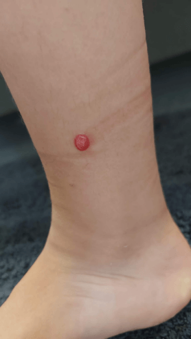
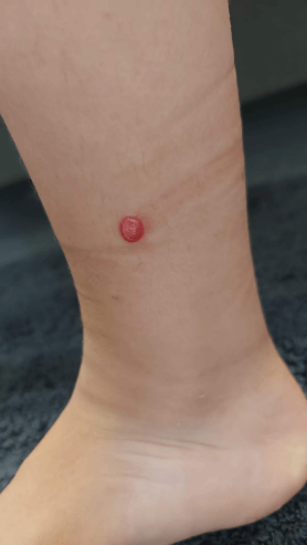
A six-year-old male child, otherwise healthy, was referred to the dermatology department for a pink papular skin lesion on the right lower limb (Figure 1), which was progressively growing over 4 months, without associated itching, bleeding, or pain. On physical examination, the lesion measured approximately 6 mm in diameter with irregular borders and elevated topography. Dermatoscopy did not show the typical starburst or peripheral globular patterns, instead revealing nonspecific vascular structures. An excisional biopsy was performed, and the histopathological report revealed a symmetric and well-demarcated compound melanocytic proliferation, predominantly composed of spindle-shaped, voluminous melanocytes. There was no atypia, and rare superficial mitotic figures were observed. Surgical excision was complete, and the margins were clear.
Figure 1. Pink papular skin lesion on the right lower limb.
Case 2
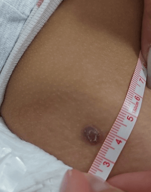
A three-year-old female child, without any significant medical history, was referred to a dermatology appointment due to the appearance of an erythematous-violaceous nodule approximately 10 mm in diameter on the right flank (Figure 2), which had been progressively growing over 5 months. On physical examination, the lesion measured 10 mm in diameter, with irregular borders, an elevated topography, and signs of ulceration. An excisional biopsy of the lesion was performed, and histopathological analysis confirmed a Spitz nevus. The lesion exhibited a symmetric architecture with sharply demarcated borders and was predominantly composed of spindle cells. Notably, the analysis revealed a rare mitotic rate, indicating very few mitotic figures in the tissue sample. This is a critical histopathological finding, as a low mitotic rate is characteristic of benign lesions and reflects minimal cellular proliferation. The absence of frequent mitoses, along with the lack of other atypical features (pleomorphism, disorganized growth, or deep tissue invasion), strongly supported the benign nature of the lesion. Consequently, these findings eliminated the need for additional molecular studies, as there was no evidence of atypia or malignancy.
Figure 2. Eerythematous-violaceous nodule on the right flank.
|
Figure 1. Pink papular skin lesion on the right lower limb.
|
What is the clinical importance of analyzing spitz nevus?
In both cases, the clinical diagnosis considered was Spitz nevus. The presentation of Spitz proliferations in children is distinctive from the adults. Benign Spitz nevus typically manifest as solitary, pink-red or reddish-brown papules or dome-shaped nodules, or dark brown to black macules or papules, usually measuring 5 to 6 mm in diameter.1,2 They typically exhibit regular, raised borders and are primarily observed on the face and lower limbs. These lesions often emerge suddenly, undergo rapid growth over a few months, and then stabilize.1,2,3 A small subset of spitzoid proliferations, known as atypical Spitz nevus, may display irregular features such as larger size (>10 mm), irregular borders, ulceration, recent or irregular growth, symptoms of itch or pain, and history of bleeding or trauma. Over time, most of Spitz nevus appear to regress or develop clinical and dermoscopic features consistent with common acquired nevus, while others remain stable.1,2,3 The clinical significance of Spitz nevus lies in their similarity to melanoma, both clinically and histologically.3 Diagnosing Spitz nevus can be challenging, especially in children presenting with new pink-red or brown papules with atypical clinical features or course. These features may suggest an atypical spitzoid proliferation or melanoma.2,3
The clinical diagnosis of Spitz nevus can often be made based on typical clinical features, onset, and course, supported by dermoscopic findings. These lesions commonly appear as pink-red or reddish-brown papules or dome-shaped nodules. Dermoscopy often reveals characteristic patterns, such as the starburst pattern, globular pattern, or symmetrically distributed dotted vessels. The starburst pattern consists of radiating streaks or projections extending symmetrically from the lesion's periphery, reflecting the radial proliferation of melanocytes and commonly seen in rapidly growing Spitz nevus. The globular pattern is characterized by evenly distributed pigmented globules or dots, corresponding to clusters of melanocytes, and is typical of stable or maturing lesions. Dotted vessels, which appear as symmetrically distributed pinpoint dots, represent dilated capillaries and contribute to the lesion’s pink or reddish hue. The symmetry and uniformity of these patterns are key features that distinguish Spitz nevus from atypical or malignant melanocytic lesions.2,4 Biopsy is recommended for lesions displaying atypical clinical and/or dermoscopic features or showing growth or changes during close interval follow-up visits.5,6 According to the modified pediatric ABCD criteria (amelanotic, bleeding, bump, uniform color, development of new, and any diameter), both described nevus warranted excisional biopsy and histopathological analysis.3,5,6 Excisional biopsy allows for the evaluation of the lesion's overall architecture and specific histological features, which are crucial for determining whether the lesion is benign or malignant.3,5,6,7
In conclusion, distinguishing Spitz nevus from malignant melanoma is crucial to avoid overtreatment and is among the most challenging issues in dermatopathology.3 In most of the cases, Spitz nevus in children are benign and have a favorable outcome.1,2,3 However, lesions with atypical clinical and dermoscopic features have a higher risk of representing melanoma, particularly in adolescents.5,6 Such lesions should be referred to dermatology and excised for histopathologic examination. |
| |
| Compliance with ethical standards |
|
Funding: None
|
|
|
Conflict of Interest: None
|
|
- Barnhill RL. The Spitzoid lesion: rethinking Spitz tumors, atypical variants, 'Spitzoid melanoma' and risk assessment. Mod Pathol 2006; 19 Suppl 2:S21. [CrossRef] [PubMed]
- Luo S, Sepehr A, Tsao H. Spitz nevi and other Spitzoid lesions part I. Background and diagnoses. J Am Acad Dermatol. 2011;65(6):1073-1084. doi:10.1016/j.jaad.2011.04.040 [CrossRef] [PubMed] [PMC free article]
- Zaenglein AL, Heintz P, Kamino H, et al. Congenital Spitz nevus clinically mimicking melanoma. J Am Acad Dermatol 2002; 47:441. [CrossRef] [PubMed]
- Diaconeasa A, Boda D, Solovan C, Enescu DM, Vîlcea AM, Zurac S. Histopathologic features of Spitzoid lesions in different age groups. Rom J Morphol Embryol. 2013;54:51-62.
- Cordoro KM, Gupta D, Frieden IJ, McCalmont T, Kashani-Sabet M. Pediatric melanoma: results of a large cohort study and proposal for modified ABCD detection criteria for children. J Am Acad Dermatol. 2013;68(6):913-925. doi:10.1016/j.jaad.2012.12.953 [CrossRef] [PubMed]
- American Academy of Dermatology (AAD) Association. Melanoma Can Look Different in Children (https://www.aad.org/public/diseases/skincancer/types/common/melanoma/different-children). Accessed 11/10/2022.
- SPITZ S. Melanomas of childhood. Am J Pathol 1948; 24:591. [CrossRef]
|
|
DOI: https://doi.org/10.7199/ped.oncall.2026.37 |
| |
Cite this article as:
Magalhães T L d, Miranda S P, Bernardo D, Azevedo A, Machado S. Spitz Nevus and its Clinical Relevance. Pediatr Oncall J. 2025 Jan 15. doi: 10.7199/ped.oncall.2026.37
|