|
Third Fontanelle - case report
Catarina Sofia Gonçalves Afonso, Maria Beatriz Ferreira Rodrigues de Morais, Daniela Figueiredo Ramos.
Pediatrics, Hospital de Vila Franca de Xira, Unidade Local de Saúde Estuário do Tejo, Vila Franca de Xira, Portugal.
ADDRESS FOR CORRESPONDENCE
Catarina Sofia Gonçalves Afonso, Estrada Carlos Lima Costa nº2, Povos, 2600-009 Vila Franca de Xira, Lisbon, Portugal.
Email: catarinasofiaafonso@gmail.com
Newborn, Anterior fontanelle, Posterior fontanelle, Third fontanelle, Sagittal/parietal fontanelle
A term newborn boy, delivered by cesarean section, presented at birth with a large anterior fontanelle (3x4 cm), a large posterior fontanelle (3x3 cm) and a defect at the junction of the sagittal suture, with parietal bones about 5mm apart. Palpation of the fontanelle in the upright position reveal a normal slight pulsation. The remaining physical examination was unremarkable, without dysmorphias or malformations. He had good tone and primitive reflexes and no neurological signs. His birth weight was 3.38 Kg (50th percentile), height was 48.5 cm (15th-50th percentile) and head circumference 36 cm (85th-97th percentile). He had no signs of hip dysplasia (Ortolani or Barlow maneuvers) or head asymmetry.
The mother is natural from Africa and has thalassemia and sickle cell trait. The first trimester ultrasound scan showed increased nuchal translucency, which led to amniocentesis - karyotype 46 XY. The mother reports no history of prenatal infection. Routine prenatal screening for syphilis was negative. She is immune to rubella and non-immune to toxoplasmosis.
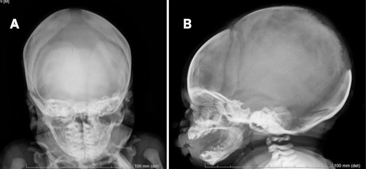
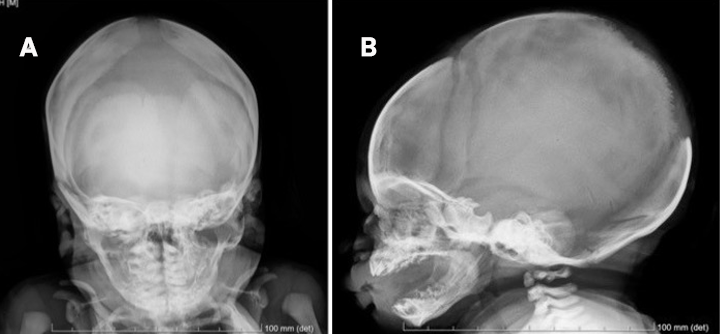
A skull X-ray performed in the coronal plane showed large anterior fontanelle and widening of the sagittal suture, and in the sagittal plane loss of continuity of skull and indentation of parietal bones near to transition parieto-occipital. (Figure 1). On coronal radiography, the anterior fontanelle measured 5.7 x 2.6 cm, and the interparietal distance was 11 mm. Cranial ultrasound showing no abnormalities, namely signs of hydrocephalus.
Complete blood count, liver, renal and thyroid function tests showed normal results. Phospho-calcium metabolism showed vitamin D deficiency (13.4 ng/ml) and slight decrease in alkaline phosphatase (99 UI/L), but parathyroid hormone, calcium and phosphorus levels were normal.
He was discharged with routine vitamin D supplementation. In the follow-up appointments he showed appropriate psychomotor development and regular head growing above 97 percentile. At six weeks of age the sagittal bone gap was closed. At 5 months the posterior fontanelle was closed and anterior fontanelle had 2x2 cm starting to calcify. At seven months the anterior fontanelle remains patent, though it is undergoing calcification without no membranous portion observed. At 13 months the anterior fontanelle was already closed.
Blood tests reassessment showed normalization of vitamin D levels, no anemia and no iron deficiency. The Mentzer index was suggestive of thalassemia and hemoglobin chromatography revealed the presence of HbS, so he was referenced to Hematology appointment, in order to keep hemoglobinopathy investigation.
Figure 1. A - Coronal section with large anterior fontanelle and widening of the sagittal suture - the anterior fontanelle measured 5.7 x 2.6 cm, and the interparietal distance was 1.1 cm.; B - Sagittal section with loss of continuity of skull and indentation of parietal bones near to transition parieto-occipital.
|
Figure 1. A – Coronal section with large anterior fontanelle and widening of the sagittal suture - the anterior fontanelle measured 5.7 x 2.6 cm, and the interparietal distance was 1.1 cm.; B – Sagittal section with loss of continuity of skull and indentation of parietal bones near to transition parieto-occipital.
|
What is the third fontanelle?
Fontanelles are the fibrous, membrane-covered gaps created when more than two cranial bones are juxtaposed, as opposed to sutures, which are narrow seams of fibrous connective tissue that separate the flat bones of the skull. In the newborn we can find six fontanelles: the anterior, at the juncture of the two parietal and two frontal bones and the bigger one; the posterior, at the junction of the occipital; two mastoids, and two sphenoids.1,5
At birth, the newborn's skull notably features an anterior fontanelle with an average size of 20 mm and a posterior fontanelle with an average size of 5 mm. They close between 4-24 months and at 2 months, respectively.1,2 Additionally, a third fontanelle may be present (prevalence 2-6%) with dimensions ranging from 7 to 30 mm. The third fontanelle, parietal/sagittal fontanelle or Gerdy’s fontanelle is not a real fontanelle but a bony defect anterior to the posterior fontanelle along the sagittal suture that is thought to be due to incomplete ossification of the parietal bones. This fontanelle typically closes by three months of age.3,4 Some factors that can affect the size of the fontanelle are gestational age, mode of delivery, gender, ethnicity and genetic predisposition. Melanodermic newborns tend to have slightly larger dimensions.
In the largest study, which included 966 full-term and 54 preterm infants, 6.3% had a third fontanelle. None of the infants in this study had associated major congenital malformations or congenital infections. However, given that a third fontanelle has been reported in children with Down syndrome, congenital infections (syphilis and rubella), skeletal disorders and hip dislocation, some authors consider it to be a potential sign of these disorders.
An abnormal fontanelle in an infant can indicate a serious medical condition. Therefore, it is important to understand the wide variation of normal.
In the presented case, a large anterior and posterior fontanelles and presence of third fontanelle in a child without any other pathological findings in detailed anamnesis and physical examination, as well in subsidiary examinations performed, allowed us to conclude that we are dealing with a normal variant, recommending only clinical surveillance of fontanelle, head growing and neurodevelopment.
Parents should be reassured and informed about this non pathologic variabilities. |
| |
| Compliance with ethical standards |
|
Funding: None
|
|
|
Conflict of Interest: None
|
|
- Kiesler J, Ricer R. The abnormal fontanel. Am Fam Physician [Internet]. 2003; 67(12):2547-52.
- Woldeyes DH, Kiros MD, Abegaz BA, Woya AA. The Size of Anterior Fontanelle and Its Determinants at Birth Among Neonates in Northern Ethiopia: A Cross-Sectional Study. Pediatr Health Med Ther [Internet]. 2020; 11:477-83. Available from: https://doi.org/10.2147/phmt.s283857 [CrossRef] [PubMed] [PMC free article]
- Ribeiro A, Ferreira J, Dias Ângela. Fontanela anterior persistente - patológico ou variante do normal?. REVNEC [Internet]. 2018; 27(1):43-5.
- Drutz JE. The pediatric physical examination: HEENT. In: Torchia MM, editor. UpToDate [Internet]. Waltham (MA): UpToDate, Inc.; 2024 [citated 2025 Abr 28]. Available from: https://www.uptodate.com/contents/the-pediatric-physical-examination-heent
- Chemke J, Robinson A. The third fontanelle. J Pediatr. 1969 Oct;75(4):617-22. doi:10.1016/S0022-3476(69)80457-9. [CrossRef] [PubMed]
|
|
DOI: https://doi.org/10.7199/ped.oncall.2027.4 |
| |
Cite this article as:
Afonso C S G, Morais M B F R d, Ramos D F. Third Fontanelle - case report. Pediatr Oncall J. 2025 Dec 16. doi: 10.7199/ped.oncall.2027.4
|