Sundari S, Kumarasamy K, Durai Arasan G, Srinivasan V, Karamath SP, Riswana F.
Department of Pediatrics, Institute of Child Health and Hospital for Children, Egmore, Chennai, India.
ADDRESS FOR CORRESPONDENCE
Dr Sundari Subramanian, Department of Pediatrics, Institute of Child Health and Hospital for Children, Egmore, Chennai, India 600008.
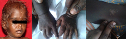
Email: drsundari.ped@gmail.com | | Keywords | | porphyria, haemolytic anemia, infancy | | | Congenital erythropoietic porphyria (CEP) is a rare metabolic disorder leading to hemolytic anemia, skin blisters, scarring, reddish urine, pigmented hair, hypertrichosis and discoloured teeth. An 11 month old male infant born of consanguineous marriage presented with abdominal distension of nine months duration with increasing pallor. There was history of passing high colored urine for 6 months. Birth history and development was normal. On examination the infant was alert, had pallor with a diffuse hyperpigmentation, sparse hypopigmented scalp hair, few dyspigmented scars and hypertrichosis (Figure 1) with splenohepatomegaly. His vital signs and anthropometry were normal. Other systems were normal. Investigations showed white cell count of 8500 cells/cumm (polymorphs 48%, lymphocytes 50%), hemoglobin of 5 gm%, and platelet count of 80,000 cells/cumm. Peripheral smear showed severe hypochromasia, anisopoikilocytosis, many polychromatic red blood cells, teardrop cells, target cells and platelets in clumps. Reticulocyte count was 4%. Liver and renal function tests were normal. HIV by Elisa, Mantoux test were negative and coomb’s test was negative. Vitamin B12 assay and hemoglobin electrophoresis were normal. Bone marrow aspiration revealed hypocellular marrow with myeloid: erythroid ratio of 3:1 and < 2% blasts and no storage cells. Urine metabolic screening was negative and stool occult blood was negative. X-ray long bones were normal. Urine did not reveal any proteins or deposits. Urine was reddish brown which changed to purplish red on standing. Cutaneous scars, hyperpigmentation, hypertrichosis, anemia and splenomegaly with erythrodontia and burgundy color urine favored the diagnosis of porphyria. Urinary porphyrins were elevated (>700mg/day). Woods lamp examination of urine and teeth showed brilliant fluorescence. Erythrocyte fluorescence testing was positive. A diagnosis of CEP was made. Confirmatory testing by enzyme assay (uroporphyrinogen synthase enzyme) was not done due to non-availability. Infant was transfused with packed red blood cells, along with supplementation of vitamin D and beta-carotene.
Figure 1: Child with depigemented hair and hyperpigmented skin
Porphyrias are disorders of biosynthesis of heme. Acute porphyrias include aminolevulinic acid dehydratase deficiency porphyria, acute intermittent porphyria, hereditary coproporphyria and variegate porphyria. Non-acute porphyrias include CEP, porphyria cutanea tarda, hepatic erythropoietic porphyria and erythropoietic protoporphyria. CEP is due to the deficiency of uroporphyrinogen III synthase enzyme. Symptoms appear in infancy as presented above. Blisters and vesicles in sun exposed areas, with scarring and deformities of the skin is common. (1) Diet, drugs, sun exposure or phototherapy precipitate the symptoms. (2) Deposits in the teeth lead to red staining and its fluorescence called as erythrodontia. Long bones may show expansion and/or mild bone loss. Diagnostic tests include urinary porphyrins (3), spectroflurometric tests, decreased enzyme activity or increased uroporphyrin1 and coproporphyrin 1 isomers and genetic studies (mutation in biallelic mutation in URO synthase or identification of the mutation in hemizygous linked gene GATA1). Treatment of CEP includes avoidance of triggers and use of sunscreens. Blood transfusions and splenectomy provide short term benefits. Activated charcoal may help in remission. Bone marrow transplant is the only cure. (4) | | | | Compliance with Ethical Standards | | Funding None | | | | Conflict of Interest None | | |
- Congenital erythropoietic porphyria – gene reviews. Downloaded from www.ncbi.nlm.nih.gov. Accessed on 21.06.2015.
- Singh DK, Ruchi R. Congenital erythropoietic porphyria. Indian Pediatr. 2008; 45:865-866. [PubMed]
- Sinharay M, Deb N, Mukhopadhyay M. Clinical laboratory approach in a case of congenital erythropoietic porphyria. Gomal J Med Sci 2012;10:253-6.
- Shaw PH, Mancini AJ, Mc Connell JP, Brown D, Kletzel M. Treatment of congenital erythropoietic porphyria in children by allogeneic stem cell transplantation: a case report and review of the literature 2001;27(1):101-105.
DOI: https://doi.org/10.7199/ped.oncall.2015.65
|
| Cite this article as: | | S S, K K, G D A, V S, SP K, F R. Congenital Erythropoietic Porphyria-An Unusual Cause of Hemolytic Anemia in an Infant. Pediatr Oncall J. 2015;12: 113-114. doi: 10.7199/ped.oncall.2015.65 |
|