Mariana Vieira da Silva1, Inês Maria Costa Andrezo Lobo1, Jorge Filipe Costa Rodrigues1, Sónia Coelho2, Maria Gabriela Mendes Pais Lopes Laranjo1, Cristina Celeste Fernandes de Faria1
1Department of Pediatrics, Centro Hospitalar Tondela-Viseu, Viseu, 3504-509, Portugal, 2Department of Dermatology, Centro Hospitalar Tondela-Viseu, Viseu, 3504-509, Portugal
Address for Correspondence: Mariana Vieira da Silva, Department of Pediatrics, Centro Hospitalar Tondela-Viseu, Rua dos passionistas, nº 130, 4º direito frente, 4520-292- Santa Maria da Feira, Portugal.
Email: vieirasmariana24@gmail.com
|
Question :A 14-month-old boy presented to the emergency department with an 8-day history of fever and oral aphthous lesions. On day 5 of illness, he was apyretic for almost 24 hours. On day 6, low-grade fever reappeared associated with nasal congestion, cough, diarrhea, facial rash and edema of both ears and amoxicillin/clavulanic acid was started given suspicion of cutaneous infection. After 48 hours, due to worsening of the rash and progressive peripheral edema, he was admitted to hospital for further investigation. No other symptoms nor history of suspicious exposures were found. Personal and family history were also unremarkable.
Physical examination showed purpuric lesions with a cockade pattern, localized to his face (cheeks and ears) and limbs. He also had significant non-pitting edema of the auricles and extremities. Multiplex PCR respiratory panel was positive for Adenovirus and Rhinorivus- Enterovirus. The remaining laboratory results and chest X-rays were normal.
The child was admitted considering the possibility of either acute hemorrhagic edema of infancy (AHEI) or post-infectious polymorphous erythema. During hospitalization, antibiotic therapy was suspended and a skin biopsy was carried out. Emollient cream and topical corticosteroid therapy were started with clinical improvement and he was discharged after 4 days. Clinical reassessment was carried out after 3 weeks, with resolution of the lesions and confirmed pathological diagnosis of AHEI.
Figure 1. Face and ear, day 8 and day 9. 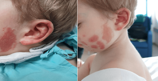 Figure 2.
 Right arm, day 8 and day 9. 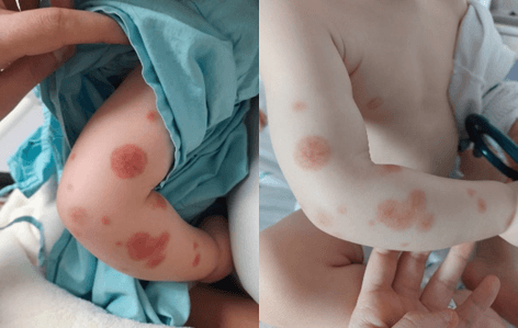 Figure 3.
 Left hand, day 8 and day 9. 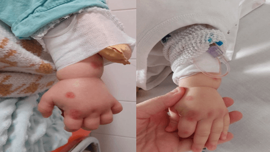 What is Acute hemorrhagic edema of infancy?
|
Discussion :
AHEI is a small-vessel vasculitis of sudden onset and unknown etiology. It is a rare condition predominantly affecting children under 24 months. Unlike IgA vasculitis (the main differential diagnosis), AHEI is rarely associated with systemic symptoms (apart from fever), has more localized lesions, normal laboratory tests and spontaneous resolution. | References : | - Caixeta MF, Lima JS, Zandonaide AGB. Edema agudo hemorrágico da infância : Relato de caso e comparação com meningococcemia. 2016;6(2):98-102.
- Garcia C, Sokolova A, Torre MDL, Amaro C. Edema agudo hemorrágico da infância. Rev Port Imunoalergologia. 2013;213-4.
- Lacerda ACM, Silva SA, Rafael MS, Correia SAM, Batista CB, Castanhinha SIF. Edema hemorrágico agudo da infância: uma vasculite com bom prognóstico. 2014;24(2):137-41.
|
|
| Correct Answers : |  100% 100% |
Last Shown : Apr 2025
|