Mafalda João Pereira, Íris Rocha e Oliveira, Andreia J. Fernandes, Maria João Virtuoso
Pediatric Department, Unidade Local de Saúde do Algarve, Hospital de Faro, Faro, Portugal
Address for Correspondence: Mafalda João Pereira, Hospital de Faro, Rua Leão Penedo, 8000-386 Faro, Portugal.
Email: mjcpereira@chalgarve.min-saude.pt
|
Question :A seven-year-old girl, with a previous history of severe atopic dermatitis (AD) and sensitization to several allergens, presented to the pediatric emergency department (ED). She was previously followed by a Pediatric Allergologist since she was six months-old and medicated daily with desloratadine and topic tacrolimus, plus topic fluticasone propionate in acute flares. She had interrupted her medication for the last two months.
She presented to the ED complaining of pruritic skin lesions, progressively worse during the past week, and gait limited by pain. She denied fever or other symptoms. On physical examination, she displayed xerosis, scaly eczematous lesions, thickening of the skin and an increase in skin markings (lichenification), with extensive and painful impetiginous lesions scattered throughout the body, worse in the lower and upper limbs and around the neck (Images).
Analytically she had an increased C-reactive protein but no leukocytosis, with eosinophils in the upper normal limit (800 cells/uL). Her blood culture was positive for methicillin-sensitive Staphylococcus aureus. Her total IgE was elevated (3340.0 kUI/L) with specific IgE positive for several types of mites and olive tree.
Treatment was initiated with flucloxacillin, daily hygiene with a chlorhexidine solution, skin hydration with application of an emollient (moisturizer) and a topic steroid (betamethasone dipropionate twice daily).
By the time of discharge, there was a clear improvement, with lesions in the healing phase, decreased pruritus, and no functional impairment. She resumed follow-up with the Pediatric Allergologist and resumed treatment with oral antihistamine, topic tacrolimus, and topic steroids in flares.
Figure 1. Infected atopic dermatitis in the back of the neck. 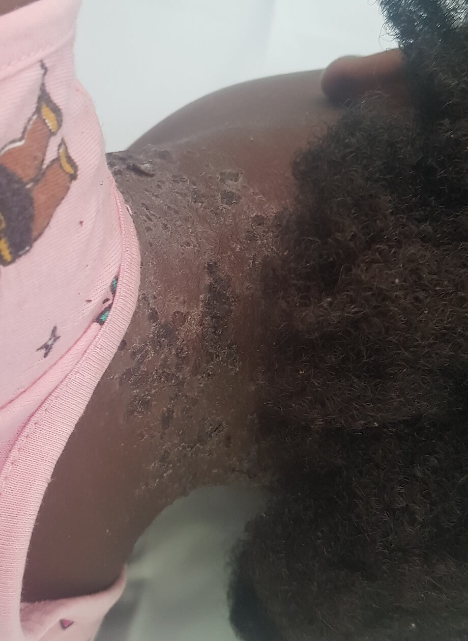 Figure 2.
 Infected atopic dermatitis in the legs. 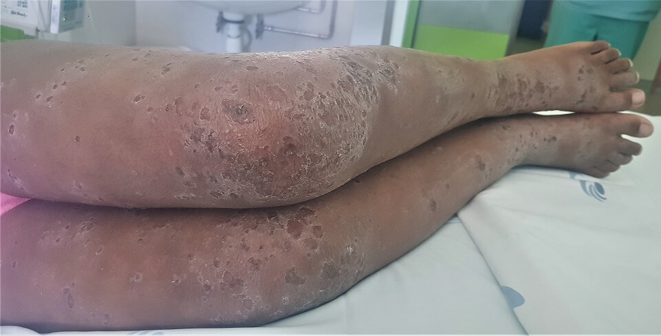 What is the diagnosis?
|
Discussion :
AD is a chronic, relapsing, and highly pruritic inflammatory skin condition, which generally develops in early childhood. 1,2,3 Five major clinical features of AD are: pruritus; a chronic, relapsing course; typical distribution; family or personal history of atopy; onset before 2 years of age. 1,2,3
Diagnosis can be difficult, since it can be mistaken for other skin conditions, such as seborrheic dermatitis, with usually less pruritic lesions, and salmon-colored patches with greasy scales; contact dermatitis, with more localized lesions and a history of a locally-applied allergen; scabies, very pruritic but palmar and plantar skin lesions and more family members affected; several primary immunodeficiency diseases, that present with eczematous rash but usually present in the first weeks-to-months of life and have several distinguishing systemic features; among other conditions. 1,2,3
Staphylococcus aureus colonization is common in AD, affecting over 90% of patients, and the density of S. aureus on the skin correlates directly with AD severity. 1,3 Optimal control of all aspects of AD morbidity, including pruritus, is best achieved through restoration of the skin barrier by adequate hydration and control of skin inflammation. 1,2| References : | - Lyons, Jonathan L, et al. Atopic Dermatitis in Children: Clinical Features, Pathophysiology and Treatment, Immunol Allergy Clin North Am. 2015 February; 35(1): 161-183. Doi: 10.1016/J.iac.2014.09.008.
- Weston, William M, and William Howe. "Atopic Dermatitis (Eczema): Pathogenesis, Clinical Manifestations, and Diagnosis." UpToDate, 30 May 2020, www.uptodate.com/contents/atopic-dermatitis-eczema-pathogenesis-clinical-manifestations-and-diagnosis?topicRef=1726.
- Deleuran, M, and C Vestergaard. Clinical heterogeneity and differential diagnosis of atopic dermatitis, British Journal of Dermatology (2014) 170 (Suppl. s1), Pp 2-6. DOI 10.1111/Bjd.12933.
|
|
| Correct Answers : |  100% 100% |
Last Shown : May 2025
|