A 6-week-old male infant was brought to the emergency department with a three-day history of irritability, decreased food intake and a polymorphic skin eruption. He had no significant medical history, was exclusively breastfed with good weight gain. None of his family members had similar symptoms or lesions. The infant was in good general condition. Skin examination revealed an erythematous base with scattered vesicles, pustules and papules. The lesions were more prominent on the scalp, back and abdomen, including the neck, inguinal and axillary regions (Figure 1). Blood parameters were within reference ranges for age, with no increased inflammatory markers. Nevertheless, due to the exuberance of the lesions, a bacterial infection was suspected and intravenous flucloxacillin was initiated.
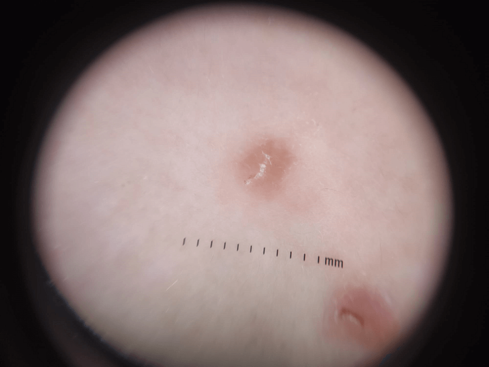
The following day, the infant presented with new lesions and nodular scabies was suspected. The delta-wing jet sign observed on direct dermoscopy confirmed the diagnosis (Figure 2). The patient and his parents were treated with topical 6% sulfur ointment for three consecutive days, repeated after seven days.
In the follow-up evaluation four days after completing treatment, the infant presented with additional lesions on his back. His aunt had been taking care of him in the previous days and neither she nor her family had been treated. The patient and all his close contacts underwent two more cycles of treatment, with complete resolution of the lesions.
Figure 1A and 1B. Lesions in the torso: scattered vesicles, pustules, papules and nodules in an erythematous base.
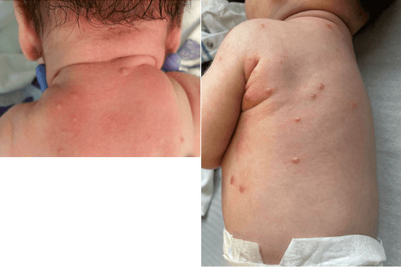 Figure 2.
 Dermoscopy of the lesions: delta-wing jet sign, a triangular structure at the end of the central S-shaped burrow.
What is the Diagnosis?