Aishwarya Padubidri Muralidhar1, Tapan Kumar Dash2
1Paediatric Cardiac ICU, CARE Hospitals, Banjara Hills, Hyderabad, 2Director and Head of Department, Paediatric CTVS department, CARE Hospitals, Banjara Hills, Hyderabad
Address for Correspondence: Aishwarya Padubidri Muralidhar, Paediatric Cardiac ICU, CARE Hospitals, Banjara hills road no 1, Hyderabad.
Email: aishu.padubidri.m@gmail.com
|
Question :A 15-month-old female child presented with a 6-month history of progressive refusal to eat solid food, intermittent choking episodes, noisy breathing and failure to gain weight. The child was born out of a non-consanguineous marriage and third in birth order. The baby was born via normal vaginal delivery at term, cried immediately after birth, weighed 3.2 kg and had a smooth perinatal transition. The child was exclusively breastfed for the first 6 months and was started on complementary feeding thereafter. Initially, the mother reported that the baby accepted the feeds well, but gradually, the baby began to refuse solid food while continuing to accept the mother's milk, with intermittent choking episodes. The child was observed to have progressively worsening noisy breathing associated with fast breathing, which increased on crying. There was no history of fever, persistent coughing, regurgitation, drooling, snoring, mouth breathing, or recurrent upper respiratory tract infection.
On physical examination, the child was found to be alert. Her anthropometry was below the 3rd percentile for height and weight. Mild pallor was noted, but no cyanosis, clubbing, or oedema was present. Respiratory examination revealed tachypnoea (respiratory rate 40/min) and audible biphasic stridor. Cardiac examination was unremarkable. Other general and systemic examinations were normal.
Chest X-ray was done which revealed an abnormally widened superior mediastinum as in Figure 1, suggesting a possible vascular ring. This was confirmed by a contrast-enhanced CT scan of the chest, which showed a double aortic arch compressing the trachea and oesophagus (Figure 2 and Figure 3). No other associated cardiac anomalies were noted. Patient underwent surgical intervention to divide the anomalous vascular ring. Postoperatively, the child was initiated on liquid diet followed by a gradual transition to solids, which were accepted without difficulty. The child was discharged home on postoperative day 6. At 6 months of follow up the child is asymptomatic and gaining weight on regular diet.
Figure 1. Chest X-ray showing widened superior mediastinum (white arrow) and prominent right aortic arch (blue arrow). 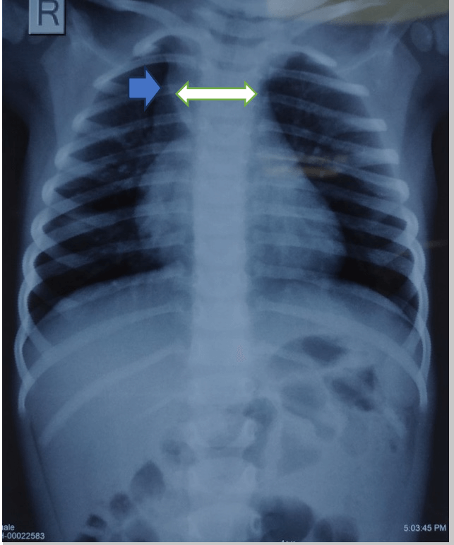 Figure 2.
 CECT chest in axial section showing Double aortic arch that is right aortic arch and left aortic arch forming a vascular ring. 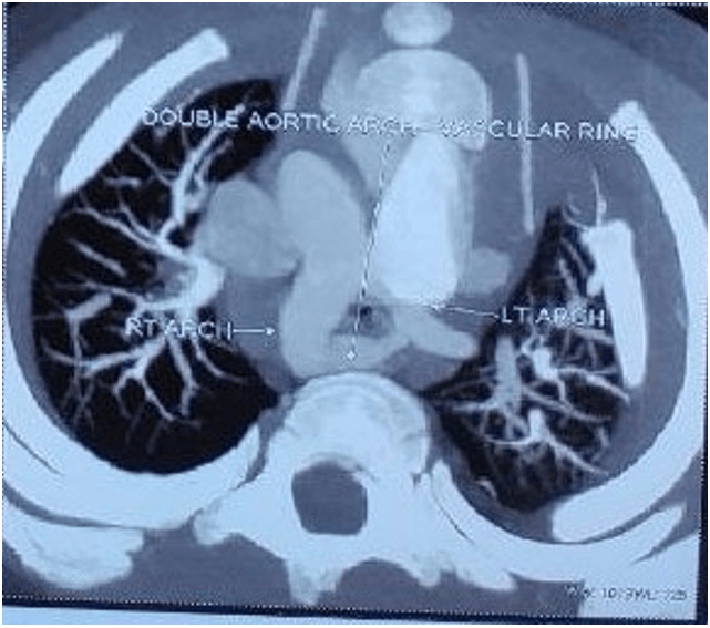 Figure 3.
 CECT chest: axial section(A), lateral section (B), oblique lateral section(C) and coronal section(D) showing compression of the trachea and oesophagus by the vascular ring. 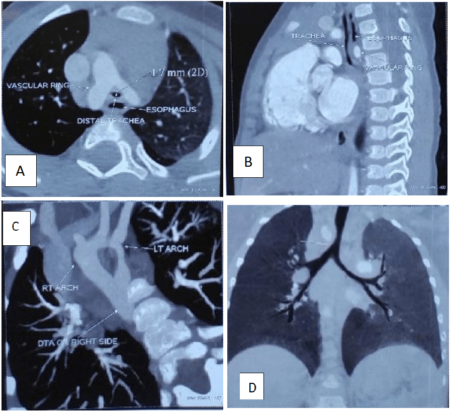 What is vascular ring?
|
Discussion :
Vascular ring is a rare vascular anomaly, comprising approximately 1% of all congenital cardiovascular abnormalities where the aorta or its branches encircle the trachea and oesophagus leading to their compression. 1 The most common type of vascular ring in symptomatic patients is a double aortic arch, which accounts for approximately 80% of cases. With improving imaging facilities, right aortic arch with left ligamentum arteriosum is increasingly being diagnosed. Other rarer types are left aortic arch with aberrant right subclavian artery, circumflex aorta and pulmonary artery sling. 2 It can present with varying degree of respiratory (90%) and gastrointestinal (50%) symptoms such as noisy breathing, seal-like barking cough, recurrent respiratory tract infections, dysphagia, slow eating, failure to thrive, at any age, from infancy to adulthood, with a subset of patients remaining asymptomatic throughout life. 3
The diagnosis of vascular ring requires high clinical suspicion and is made by imaging studies. Chest X-ray may show a wide superior mediastinum or a right-sided aortic arch. Contrast enhanced CT scan is preferred for diagnosis and can provide detailed information about the anatomy of the vascular ring and degree of compression of the trachea and oesophagus. Additional information may be obtained with CT angiography and 3D reconstruction images. Associated structural cardiac anomalies may be found in 12% of the patients and hence echo should be included in the workup. 4
The treatment of symptomatic vascular ring is surgical. The goal of surgery is to divide the aberrant blood vessels that are encircling the trachea and/or oesophagus which can be done by thoracotomy or minimally invasive methods. The prognosis for patients with vascular ring is excellent after surgical correction. The management of asymptomatic and incidentally detected vascular rings includes close follow up and serial imaging to decide on intervention. 5
This case highlights the importance of considering vascular ring as a differential diagnosis in infants presenting with feeding difficulties, stridor and failure to thrive. Early diagnosis and surgical intervention are crucial for optimal outcomes. | References : | - Bonnard A, Auber F, Fourcade L,et al. Vascular ring abnormalities: a retrospective study of 62 cases. Journal of pediatric surgery. 2003 Apr 1;38(4):539-43.
- Backer CL, Mongé MC, Popescu AR, et al. Vascular rings. Semin Pediatr Surg. 2016;25(3):165-175.
- Suh YJ, Kim GB, Kwon BS,et al. Clinical course of vascular rings and risk factors associated with mortality. Korean Circ J. 2012 Apr;42(4):252-8.
- Worhunsky DJ, Levy BE, Stephens EH, Backer CL. Vascular rings. Semin Pediatr Surg. 2021;30(6):151128.
- Sahu A, Moe TG. Identification and Management of Vascular Rings and Slings. JACC Case Rep. 2024 Apr 17;29(8):102316.
|
|
| Correct Answers : |  100% 100% |
Last Shown : Aug 2026
|