Catarina Martins, Inês Vivas, Maria José Dinis.
Pediatrics and Neonatology Department, Unidade Local de Saúde de Póvoa de Varzim/Vila do Conde, Portugal.
ADDRESS FOR CORRESPONDENCE
Catarina Martins; Largo da Misericórdia, 4490-421, Póvoa de Varzim, Portugal.
Email: catarinaguimm@gmail.com | | Abstract | Background: Cutaneous mastocytosis (CM) is a rare disorder characterised by abnormal accumulation of mast cells in the skin. In children, it is usually confined to the skin and has a favourable prognosis.
Case presentation: A healthy 6-month-old male infant developed scattered erythematous macular lesions on the trunk and limbs during hospitalisation for bronchiolitis. The lesions were non-pruritic, blanched with pressure, and spared the palms, soles, genital area, and face. At one-month follow-up, the lesions persisted, appearing hyperchromic, with new lesions on the face. There was still no pruritus, elevation, or involvement of palms, soles, or genitalia, although lesions became redder after bathing. Abdominal ultrasound and laboratory work-up, including IgE and baseline tryptase, were normal. Dermatology referral and skin biopsy confirmed cutaneous mastocytosis. The infant remains under regular follow-up in general paediatrics and dermatology.
Discussion: The presentation-onset in infancy, macular lesions on trunk and limbs, sparing of palms and soles, histological confirmation, and normal systemic evaluation-is consistent with maculopapular cutaneous mastocytosis (urticaria pigmentosa), the most common paediatric form. The absence of systemic involvement indicates a benign course. Reported risk factors for systemic disease include older age at onset, persistence beyond puberty, elevated basal tryptase, and hepatosplenomegaly or lymphadenopathy.
Conclusion: Early identification of CM in infancy allows appropriate diagnosis, prevents unnecessary investigations, and guides follow-up. This case highlights that infants with typical cutaneous findings and normal systemic work-up can usually be managed conservatively with regular clinical monitoring and family reassurance regarding the favourable prognosis.
| | | | Keywords | | Mastocytosis, Cutaneous, Child | | | | Introduction | | Mastocytosis denotes a heterogeneous group of disorders characterised by abnormal accumulation and activation of mast cells in one or more organs.1 In children, the cutaneous form (cutaneous mastocytosis, CM) predominates, whereas systemic mastocytosis (SM) is uncommon. According to current classification, CM in childhood may present as maculopapular cutaneous mastocytosis (MPCM, formerly “urticaria pigmentosa”), isolated mastocytoma(s) or diffuse cutaneous mastocytosis (DCM).1 Clinical features in paediatric CM include red-brown macules or papules often on the trunk and limbs, sometimes positive Darier’s sign (urtication on rubbing) and variable symptoms due to mast cell mediator release (pruritus, flushing, blistering). Most cases begin within the first two years of life and the prognosis is generally good - many cases regress in childhood or adolescence, and progression to systemic disease is rare, but should be considered especially in presence of risk factors.2,3 Here we present a case of infantile onset CM in a healthy child hospitalised for bronchiolitis, with typical skin findings and normal systemic work-up. | | | | Case Report | A 6-month-old male infant, previously healthy and born at term, was admitted for bronchiolitis. During hospitalisation, multiple erythematous macular lesions were noted scattered across the trunk and limbs. The lesions were non-pruritic, macular, and blanching on digital pressure. They spared the palms, soles, genital area and face. No systemic symptoms - such as flushing, blistering, gastrointestinal symptoms or anaphylaxis - were observed. At discharge, the lesions were documented and the infant was referred for dermatology follow-up.
At one-month follow-up, the cutaneous lesions persisted. Many had become hyperchromic and new lesions were noted on the face. Still, the palms, soles and genital area remained uninvolved. The child had no pruritus or elevation of the lesions, but the parents noted that during bathing the lesions appeared more erythematous.
Laboratory work-up included complete blood count, liver and renal function tests, IgE level and baseline serum tryptase level. All results were within normal limits. An abdominal ultrasound was normal, with no evidence of hepatosplenomegaly, lymphadenopathy or other organ involvement. Based on the clinical presentation and normal systemic investigations, a provisional diagnosis of CM was made. A skin biopsy was performed showing dermal mast cell infiltration consistent with mastocytosis. The child was referred to dermatology for ongoing surveillance. At present (ongoing follow-up under general paediatrics and dermatology) the lesions remai, but the infant is well, with no systemic involvement or complications.
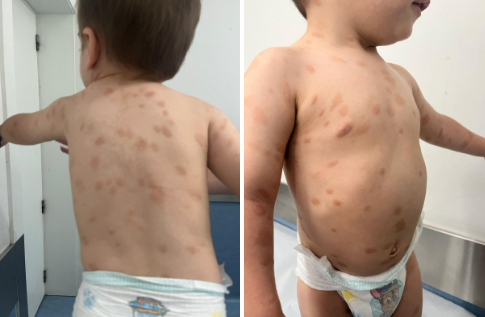
Figure 1. Erythematous macular lesions scattered across the trunk and limbs, compatible with cutaneous mastocytosis.
 | | | | Discussion | The presented case of early‐infancy onset macular skin lesions, benign course, normal systemic work-up and biopsy‐confirmed mast cell infiltration aligns with the typical presentation of paediatric CM, particularly the MPCM variant.4 Generally, paediatric CM onset typically occurs in early infancy, with trunk/limbs distribution and spare of palms/soles and genitalia (unless more extensive disease is present), with favourable prognosis in most cases.2 The onset within the first year of life, the macular non-elevated blanching lesions, absence of pruritus or systemic symptoms in this case all point to a mild phenotype.
Laboratory assessment in paediatric CM frequently reveals normal baseline tryptase and no evidence of systemic involvement in children. Elevation of serum tryptase and high number of skin lesions have been associated with increased risk of systemic, where higher lesion count, higher tryptase and more skin signs/symptoms correlate with systemic involvement.5 Thus the normal tryptase and absence of systemic features in our patient are reassuring.
Management of paediatric CM is largely conservative: reassurance, avoidance of known triggers (e.g., heat, friction, certain medications, insect stings) and symptomatic treatment (e.g., H1 antihistamines, topical therapies) when needed. Progression to systemic disease is rare, but should be monitored.3,6 Risk factors for monitoring and potential systemic disease include late onset of lesions (after age 2 years), persistence beyond puberty, presence of hepatosplenomegaly/lymphadenopathy, elevated tryptase >20 ng/m or extracutaneous mast cell infiltration.3 In our patient, none of these risk factors are currently present.
Our case had some atypical features: the absence of pruritus (many children do have pruritus, blistering or urticaria) and the sparing of palms/soles/face initially, though new lesions on the face appeared later. The blanching nature of the lesions is consistent with mast cell-mediator release. The fact that lesions became more erythematous during bathing suggests a trigger effect (heat/water). The decision to perform skin biopsy was appropriate given the need to confirm diagnosis and exclude other conditions in infancy that may mimic the presentation (e.g., pigmentary disorders, urticaria, vasculitic lesions). The normal abdominal ultrasound and labs increased confidence in limited cutaneous disease.
Long-term follow-up in our patient is recommended, particularly monitoring for any systemic signs (hepatosplenomegaly, lymphadenopathy, gastrointestinal symptoms, unexplained anaphylaxis) and periodic reassessment of skin lesion burden and tryptase levels if clinically indicated.6 Parental counselling on trigger avoidance and symptom recognition (e.g., flushing, blistering, anaphylaxis) is key. | | | | Conclusion | | In summary, we report a case of infant‐onset cutaneous mastocytosis with classical features, normal systemic evaluation and favourable initial course. Awareness of the condition among paediatricians and dermatologists is important to enable early recognition, appropriate work-up to exclude systemic involvement, and avoidance of unnecessary interventions. This case underlines the typical benign nature of paediatric CM and reinforces the value of coordinated follow-up and family education. | | | | Compliance with Ethical Standards | | Funding None | | | | Conflict of Interest None | | |
- Brockow K, Bent RK, Schneider S, Spies S, Kranen K, Hindelang B, et al. Challenges in the diagnosis of cutaneous mastocytosis. Diagnostics (Basel). 2024;14(2):161. doi:10.3390/diagnostics14020161. [CrossRef] [PubMed] [PMC free article]
- Ben-Amitai D, Metzker A, Cohen HA. Pediatric cutaneous mastocytosis: a review of 180 patients. Isr Med Assoc J. 2005;7(5):320-2. PMID:15909466.
- Sandru F, Petca RC, Costescu M, Dumitrașcu MC, Popa A, Petca A, et al. Cutaneous mastocytosis in childhood-update from the literature. J Clin Med. 2021;10(7):1474. doi:10.3390/jcm10071474. [CrossRef] [PubMed] [PMC free article]
- Hussain SH. Pediatric mastocytosis. Curr Opin Pediatr. 2020;32(4):531-8. doi:10.1097/MOP.0000000000000922. PMID:32692050. [CrossRef] [PubMed]
- Siebenhaar F, Weller K, Blume-Peytavi U, Maurer M. Mastozytosen im Kindesalter [Childhood-onset mastocytosis]. Hautarzt. 2012;63(2):104-9. doi:10.1007/s00105-011-2201-2. PMID:22290278. [CrossRef] [PubMed]
- Van Rymenam A, Sacré JP, Dezfoulian B, Dresse MF, Seghaye MC. Cas clinique: La mastocytose cutanée pédiatrique [Pediatric cutaneous mastocytosis]. Rev Med Liege. 2020;75(10):636-8. PMID:33030837.
DOI: https://doi.org/10.7199/ped.oncall.2026.81
|
| Cite this article as: | | Martins C, Vivas I, Dinis M J. Cutaneous Mastocytosis. Pediatr Oncall J. 2025 Dec 31. doi: 10.7199/ped.oncall.2026.81 |
|