Majocchi's Granuloma: A Rare Form of Tinea Corporis in an Immunocompetent Adolescent
|
|
Majocchi's Granuloma: A Rare Form of Tinea Corporis in an Immunocompetent Adolescent
14/05/2025
14/05/2025
https://www.pediatriconcall.com/Journal/images/journal_cover.jpg
Ariana Tavares1, Inês Azevedo2, Miguel Costa2, Diogo Teixeira3.
1Department of Pediatrics, Unidade Local de Saúde Gaia/ Espinho in Vila Nova de Gaia, Portugal,
2Department of Pediatrics, Unidade Local de Saúde Entre Douro e Vouga in Santa Maria da Feira, Portugal,
3Department of Dermatology, Unidade Local de Saúde Gaia/ Espinho in Vila Nova de Gaia, Portugal.
ADDRESS FOR CORRESPONDENCE
Rua Conceição Fernandes S/N, 4434-502 Vila Nova de Gaia.
Email: ariana_tavares@hotmail.com
Majocchi Granuloma, Dermatophytosis, Terbinafine, Majocchi's Granuloma
|
Clinical Problem
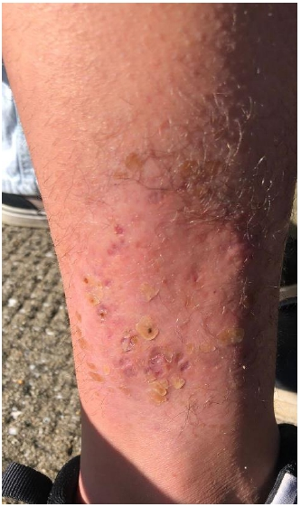
A 17-year-old female adolescent, previously healthy, was evaluated in pediatric consultation for an erythematous desquamative lesion on the lateral aspect of the right leg, associated with pruritus and pain, with approximately 1 month of evolution. She had previously received systemic corticosteroids and topical antifungal treatment without improvement and subsequently, amoxicillin with clavulanic acid without resolution. Physical examination revealed an erythematous-violaceous plaque with exudative crust and purulent drainage (Figure 1). She was referred to dermatology consultation where a skin biopsy was performed. Histopathological examination showed marked lymphohistiocytic inflammatory infiltrate with abundant neutrophils in the deep dermis and hypodermis. Mycological examination confirmed growth of Trichophytum mentagrophytes, leading to the diagnosis of tinea corporis variant Majocchi's Granuloma (MG). Treatment was initiated with terbinafine 250 mg once daily orally and ciclopirox cream for 6 weeks, with progressive resolution of the lesion. Three months after the end of therapy, only a scar lesion remained (Figure 2).
Figure 1. Erythematoviolaceous plaque with exudative crust and purulent drainage prior to treatment.
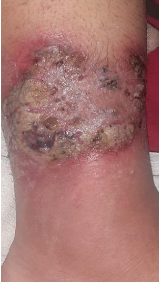
Figure 2. Scar lesion after antifungal treatment.
|
| |
When should Majocchi's Granuloma be suspected instead of other inflammatory or infectious dermatoses?
|
|
|
Discussion
MG should be considered in the presence of localized lesions that resemble superficial mycosis or eczema but are resistant to correctly administered specific topical treatments.1 It is a rare cutaneous infection caused mainly by the dermatophyte Trichophyton rubrum,3 as well as by T. violaceum and T. mentagrophytes which, instead of remaining confined to the stratum corneum, penetrate deeply along the hair follicle, extending to the dermis and hypodermis, manifesting as suppurative folliculitis with granulomatous inflammatory response. Clinically, MG is characterized by a localized area with erythematous or hyperpigmented perifollicular papules, small subcutaneous nodules or plaques and occasionally pustules. These lesions are typically refractory to conventional topical antifungal therapy due to the deep location of the fungal elements. The local immune response attempts to contain the infectious agent, resulting in the formation of characteristic granulomas, composed of histiocytes, multinucleated giant cells and mixed inflammatory infiltrate.2 Thus, the treatment of MG in immunocompetent patients necessarily requires systemic antifungal agents, such as terbinafine or itraconazole, administered for a period of 4 to 6 weeks. Oral terbinafine is the first-line treatment, as it is widely distributed throughout the body, particularly in adipose tissue. It diffuses rapidly into the dermis and accumulates in the lipophilic stratum corneum.1,3 It is essential to understand that isolated topical treatment is invariably ineffective due to the inability to reach the deep dermal structures and follicular compartments where dermatophytes establish themselves. Factors such as frequent hair removal, initial misdiagnosis (frequently interpreted as bacterial infection) and the consequent inappropriate application of topical corticosteroids significantly contribute to the exacerbation of the pathology. Corticosteroids, by inducing local immunosuppression, facilitate fungal proliferation and invasion of deeper areas of the hair follicle, deep dermis and hypodermis.3
When adequately treated, the prognosis of MG is generally promising, with complete resolution of lesions in most cases. Recurrences are relatively rare but may occur if predisposing factors (such as repeated trauma or inadequate corticosteroid therapy) are not identified and corrected.
The authors emphasize the need to avoid applying topical corticosteroids in dermatoses where the underlying infectious pathology cannot be safely excluded.4 Furthermore, they reinforce the need to consider MG as a differential diagnosis, even in patients previously treated with topical antifungals without a satisfactory response.
|
| |
| Compliance with ethical standards |
|
Funding: None
|
|
|
Conflict of Interest: None
|
- Akpadjan F, Kitha P, Dotsop L, et al. Majocchi's granuloma: an atypical clinical presentation in a child. Arch Dermatol Skin Care. 2025;7(1):6-9. Doi: 10.22259/2638-4914.0701002. [CrossRef]
- Castellanos J, Guillén-Flórez A, Valencia-Herrera A, et al. Unusual inflammatory tinea infections: Majocchi's granuloma and deep/systemic dermatophytosis. J Fungi. 2021; 7:929. Doi: 10.3390/jof7110929. [CrossRef] [PubMed] [PMC free article]
- Khodadadi RB, Yetmar ZA, Montagnon CM. Majocchi's granuloma - A multicenter retrospective cohort study. JAAD Int. 2023; 13:104-11. Doi: 10.1016/j.jdin.2023.08.010. [CrossRef] [PubMed] [PMC free article]
- Drivenes JL, Ramsing M, Bygum A. Majocchi's Granuloma - The Great Mimicker: A Case Report. Case Rep Dermatol. 2023. 18;15(1):190-193. Doi: 10.1159/000533475. [CrossRef] [PubMed] [PMC free article]
|
|
| |
Cite this article as:
Tavares A, Azevedo I, Costa M, Teixeira D. Majocchi's Granuloma: A Rare Form of Tinea Corporis in an Immunocompetent Adolescent. Pediatr Oncall J. 2025 May 13. doi: 10.7199/ped.oncall.2026.27
|