Denise Carina Chada Banganho1, Ana Filipa Ribeirinho Leal Veiga Durão2
1Serviço de Pediatria Médica, Hospital de São Bernardo, Centro Hospitalar de Setúbal, E.P.E, Setúbal, Portugal, 2Departamento de Pediatria, Hospital de Santa Maria, Centro Hospitalar e Universitário de Lisboa Norte EPE, Lisboa, Portugal
Address for Correspondence: Denise Banganho, Rua Camilo Castelo Branco, Aptd. 140, Setúbal.
Email: denise_banganho@hotmail.com
|
Question :A 29-month-old boy presented to the emergency department with a 48-hour history of left palpebral swelling without inflammatory signs or other associated symptoms. No precipitating factors were identified. The parents reported three previous similar episodes in the last 1.5 years with spontaneous resolution within 48-72 hours. The other episodes were associated with serous drainage. On examination (Figure 1) there was a left palpebral soft swelling with extension to the frontal region. In the middle of the tumefaction there was a one-millimeter orifice, without spontaneous or by expression drainage. A soft tissues ultrasound was performed at the Emergency Department, that revealed a fistulous path from clinically evident dermal lesion with apparently intracranial extension, without signs of local complications. A magnetic resonance imaging (MRI) corroborated the ultrasound findings (Figure 2). In the present case, the patient was discharged with a programed neurosurgery appointment. The boy has maintained regular follow-up in pediatric and neurosurgery consultations, waiting for spontaneous resolution, once there were no major complications in the two-year follow-up period.
Figure 1. Lateral view from the head swelling. 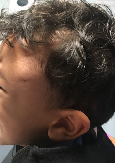 Figure 2.
 MRI image from the lesion. 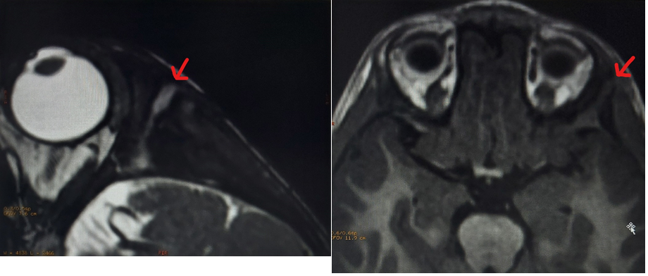 What is the diagnosis?
|
Discussion :
Orbitofacial masses represent a heterogeneous group of congenital or acquired lesions that can have inflammatory, infectious, neoplastic or traumatic etiology. 1 The most common presentation is an isolated mass with an indolent growing pattern. Some cases are associated with previous local infection. 1,2 A dermoid cyst is a cystic teratoma that contains mixed skin, hair and/or sweat gland tissue. They usually present as isolated, superficial, painless, well-circumscribed and slow-growing masses. 1 The most frequent location is the head and neck region (84% of the cases). Suppuration is uncommon. A superficial sinus orifice may be associated with an intracranial fistula. Recurring inflammatory and exudative lesions have also been described. The management of these cases is controversial: vigilance can be performed but surgical excision is the definitive treatment. 1,2
LEARNING POINTS
• Epidermoid cyst is usually asymptomatic with indolent growth.
• The presence of sinus orifice may be associated with intracranial fistula that can lead increased risks like infection.
• In the presence of a frontal mass it’s important to exclude the presence of intracranial fistula. | References : | - Stelle M, Suskind D, Moses M, et al. Orbitofacial Massses in Children. Arch Oolaryngol Head Neck Surg. 2002;128:409-413.
- Van C, Low D. A rare presentation of a dermoid cyst with draining sinus in a child: case report and literature review. Pediatric Dermatology. 2016; 33:244-248.
|
|
| Correct Answers : |  100% 100% |
Last Shown : Aug 2023
|