Reactive Infectious Mucocutaneous Eruptions: Diagnostic Challenges
Sofia Maria da Silva Faria1, Ana Rita Barroca de Macedo1, Nuno Miguel Sanches de Almeida2, Ana Luísa Figueira de Sousa Correia1, Rui Manuel Correia de Almeida1
1Pediatrics Department, Hospital Pedro Hispano, Unidade Local de Saúde de Matosinhos, Matosinhos, Portugal, 2Pediatrics Department, Centro Hospitalar de Vila Nova de Gaia-Espinho, Unidade Local de Saúde de Gaia/Espinho, Vila Nova de Gaia, Portugal
Address for Correspondence: Sofia Maria da Silva Faria, Unidade Local de Saúde de Matosinhos, Hospital Pedro Hispano, Rua de Dr. Eduardo Torres, 4464 - 513 Senhora da Hora, Portugal.
Email: sofiasilvafaria@gmail.com
Keywords: Mycoplasma pneumoniae, Human metapneumovirus, Mycoplasma pneumoniae induced rash and mucositis, reactive infectious mucocutaneous eruptions (rime), Pediatric mucositis.
Clinical Problem :
An 11-year-old girl with a past medical history of stomatitis three months earlier, presented to the emergency department with five days of mouth pain, partial food refusal, genital burning, and eye itching. She also had a productive cough for the past ten days.
On the first day of mouth pain, she was diagnosed with aphthous stomatitis at the emergency department and given supportive treatment (pain management, frequent mouth hygiene, hydration and mucosal care). Forty-eight hours later, due to the persistency of symptoms and complete food refusal, she was evaluated by her attending pediatrician, who prescribed topical betamethasone, oral Acyclovir (20 mg/Kg/dose every 6 hours), oral Azithromycin (10 mg/Kg/dose, once a day), and topical hyaluronic acid.
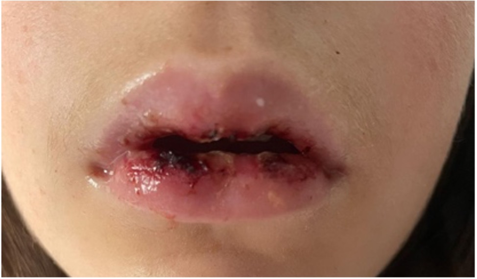
Her condition worsened, and she returned to the emergency department on the fifth day of mouth pain. Upon admission, she had no fever. Physical examination revealed hemorrhagic crusts on her lips (Figure 1), gingivitis, sloughing of the lips and tongue, and conjunctival hyperemia in her left eye, but no other skin lesions, including on her genitals.
A complete blood analysis and urine test were performed, both showing no significant abnormalities. A chest radiography was also done and showed a bronchovascular reinforcement. Additionally, multiplex Polymerase Chain Reaction (PCR) panel testing for respiratory viruses was negative for SARS-CoV-2, influenza A and B, and respiratory syncytial virus. The on-call ophthalmologist diagnosed blepharitis and superior tarsal conjunctival ulcers in her left eye. Due to complete food refusal, intravenous fluid therapy was implemented, and she was hospitalized in our Pediatric facility.
During inpatient care, an expanded multiplex PCR panel for identification of respiratory pathogens tested positive for human metapneumovirus and serologic tests were collected at admission.
Due to RIME suspicion, dermatology was consulted. Skin swab samples were collected and analyzed via PCR, which were negative for Herpes simplex type 1 and 2, Mycoplasma pneumoniae, and Chlamydia pneumoniae. Serologic tests were positive for immunoglobulin G for Herpes simplex type 1, positive for immunoglobulin M and G and for Mycoplasma pneumoniae, and equivocal immunoglobulin M but positive immunoglobulin G for Chlamydia pneumoniae.
She completed seven days of oral acyclovir, five days of oral azithromycin, four days of oral Betamethasone seven days of oral sucralfate, seven days of topical Fusidic acid and eight days of ophthalmic ointment (containing prednisolone, neomycin, and sulfacetamide).
Within a week, there was noticeable clinical improvement, with healing of the mouth ulcers, pain relief, as well as increased appetite. She was discharged home at the seventh day of hospitalization, in excellent condition. She was posteriorly followed up in a pediatric consultation and was discharged after 9 months without new episodes. No blood tests were performed.
Figure 1. Hemorrhagic crusting of upper and lower mucosal lips.
|
What is the significance of detecting human metapneumovirus in this patient, and how does it support the diagnosis of RIME over other infectious mucocutaneous conditions?
Discussion :
RIME is commonly triggered by bacterial or viral respiratory infections, with M. pneumoniae being the most common etiological agent. 1 Aside from M. pneumoniae, other pathogens that can also cause RIME are Chlamydia pneumoniae, human metapneumovirus, rhinovirus, enterovirus, parainfluenza virus 2, influenza B virus and SARS-CoV-2. 3,4,5
RIME typically presents with significant mucosal involvement, affecting multiple mucous membranes with minimal or no skin involvement. This condition, primarily affects children and adolescents, with a male predilection. The lesions can be vesiculobullous, targetoid, atypical targets, or macules. It has been previously termed M. pneumoniae-induced rash and mucositis, M. pneumoniae-associated mucositis, atypical Stevens-Johnson syndrome, or Fuchs syndrome. 1,2
Recurrence of RIME is common, and in many cases, it is triggered by a different agent than the one that caused the initial episode. 6 According to the literature, RIME recurrences tend to be less aggressive than the first episode. 6 In our patient’s case, the recurrence of RIME was more severe than the initial episode.
In the present case, the authors consider plausible that the first episode was caused by Mycoplasma pneumoniae , with the recurrence associated with the documented infection by human metapneumovirus. Co-infection by Chlamydia pneumoniae was also considered, although diagnostic tests by PCR were negative for this agent and positive serologies could be a false positive, as this is an unreliable method for its diagnosis.
There is no standard treatment for RIME. However, according to the literature, most patients are treated with antibiotic and systemic corticosteroids. A small number (8%) receive immunoglobulin. Supportive care includes pain management, hydration and mucosal care. 1,7 If eye involvement occurs, close monitoring by an ophthalmologist is essential. Treatment for conjunctivitis may include topical corticosteroid and antibiotic eye drops. 8| References : | - Waites KB, Xiao L, Liu Y et al. Mycoplasma pneumoniae from the respiratory tract and beyond. Clin Microbiol Rev. 2017;30(3):747-809.
- Valle J, Nasrollahi F, Eilbert W. Mycoplasma pneumoniae-induced rash and mucositis. Am J Emerg Med. 2022;54:324-5.
- Mayor-Ibarguren A, Feito-Rodriguez M, González-Ramos J et al. Mucositis secondary to Chlamydia pneumoniae infection: expanding the Mycoplasma pneumoniae-induced rash and mucositis concept. Pediatr Dermatol. 2017;34(4):465-72.
- Brazel D, Kulp B, Bautista G et al. Rash and mucositis associated with Mycoplasma pneumoniae and Chlamydophila pneumoniae: a recurrence of MIRM?. J Pediatric Infect Dis Soc. 2021;10(3):220-4.
- Kim JY, Reddy S, Sosa VB et al. Recurrent reactive infectious mucocutaneous eruption (RIME) caused by human metapneumovirus. J Am Acad Dermatol. 2021;85(3):98.
- Song A, Nicholson C, Maguiness S. Recurrent reactive infectious mucocutaneous eruption (RIME) in two adolescents triggered by several distinct pathogens including SARS-CoV-2 and influenza A. Pediatr Dermatol. 2021;38(5):1222-5.
- Liakos W, Xu A, Finelt N. Clinical features of recurrent Mycoplasma pneumoniae-induced rash and mucositis. Pediatr Dermatol. 2021;38(1):154-8.
- Ramien ML. Reactive infectious mucocutaneous eruption: Mycoplasma pneumoniae-induced rash and mucositis and other parainfectious eruptions. Clin Exp Dermatol. 2021;46(3):420-9.
|
| Correct Answers : |  0% 0% |
|
|
|
|
|
|