Miliary Shadows in Pulmonary Tuberculosis: How soon can they Develop?
Dhruv Gandhi, Ira Shah
Department of Pediatric Infectious Diseases, BJ Wadia Hospital for Children, Mumbai, India
Address for Correspondence: Dhruv Gandhi, 5B/13 Shyam Niwas, Breach Candy, Mumbai-400026, Maharashtra, India.
Email: dhruvgandhi2610@gmail.com
Keywords: Chest X-ray, Chest CT, Disseminated tuberculosis, Pediatric tuberculosis, Radiological diagnosis of tuberculosis, TB diagnosis.
Clinical Problem :
An 11-year-old girl presented in November 2024 with fever and dry cough for 2 days. There was no loss of weight or appetite. She was treated for microbiologically-diagnosed multidrug-resistant (MDR) miliary, abdominal and central nervous system tuberculosis (TB) 3 years ago for which she received 18 months of second-line antitubercular therapy (ATT). On presentation, her weight was 27.3kg (between 10th-25th percentile according to the Indian Academy of Pediatrics growth charts). General and systemic examinations were normal. Peripheral smear for malaria and Widal test were negative. Other investigations are shown in Table 1. She was started empirically on oral amoxicillin-clavulanate, however, there was no symptomatic improvement after 5 days of therapy. Chest X-ray performed in November 2024 showed right lower zone opacities and obliteration of the right costophrenic angle suggestive of a syn-pneumonic effusion (Figure 1A), and mycoplasma IgM was positive with a titre of 12 units/mL (normal: <10 units/mL). She was shifted onto oral Clarithromycin for 14 days during which her cough improved, however, fever spikes were persistent. On day 14 of clarithromycin, chest ultrasound showed a loculated mild right-sided pleural effusion with septations measuring about 5mL, and a thickened pleura measuring 2mm. She was started on oral levofloxacin, following which the fever spikes reduced. However, even after 14 days of levofloxacin, the fever did not subside completely and she developed a new-onset backache. Chest X-ray performed in December 2024 showed bilateral miliary pulmonary opacities (Figure 1B). Contrast-enhanced computerized tomography (CT) chest showed miliary pulmonary nodules, mediastinal lymphadenopathy, and T10-T11 vertebral end-plate destruction with periosseous collections. Gastric lavage Xpert MTB/Rif detected rifampicin-resistant Mycobacterium tuberculosis (MTB) and Xpert MTB/XDR showed resistance to Isoniazid and fluoroquinolones. She was diagnosed with pre-extensively drug-resistant TB (preXDR-TB) and was started on second-line ATT.
Figure 1. (A) Chest X-ray PA view done in November 2024 showing opacities in the right lower zone and obliteration of the right costophrenic angle. (B) Chest X-ray PA view done in December 2024 showing bilateral miliary pulmonary shadows.
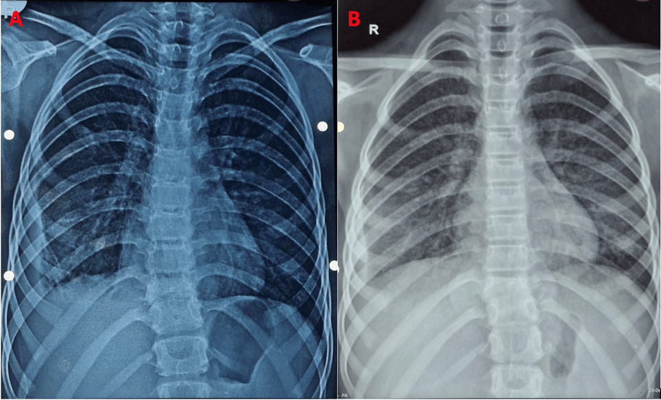
Table 1. Investigations of the patient.
| Parameters |
At presentation |
Day 14 of clarithromycin |
Day 10 of levofloxacin |
Start of ATT |
Reference Ranges |
| Hemoglobin (gm/dL) |
11.2 |
10.5 |
10.5 |
10.5 |
11.5-15.5 |
| White blood cell count (cells/cumm) |
5400 |
5480 |
5180 |
6290 |
5000-13,000 |
| Absolute neutrophil count (cells/cumm) |
2970 |
- |
3212 |
3460 |
2000-8000 |
| Absolute lymphocyte count (cells/cumm) |
2200 |
- |
1502 |
2246 |
1000-5000 |
| Platelets (105 cells/cumm) |
3.50 |
3.76 |
3.26 |
3.78 |
1.50-4.50 |
| ESR (mm/hr) |
10 |
58 |
45 |
- |
0-10 |
| CRP (mg/dL) |
- |
9.3 |
11.7 |
- |
0.3-1 |
Note: ESR- Erythrocyte sedimentation rate, CRP- C-reactive protein.
|
How soon can miliary shadows develop in pulmonary TB?
|
|
|
|
|