V Poovazhagi, Ezhilarasi S, Thangam Menon, Naveen kumar V, Chamundeeswari, Mohamed Meeran.
Pediatric Intensive Care Unit, Institute of Child Health and Hospital for Children, Egmore, Chennai.
ADDRESS FOR CORRESPONDENCE
Dr.V.Poovazhagi, 8/11 Manjolai street, Kalaimagal Nagar, Ekkaduthangal, Chennai, TamilNadu, India 600 032.
Email: poomuthu@yahoo.com | | Keywords | | Submersion injury, aspergillosis, nocardiosis. | | | Invasive aspergillosis and nocardiosis is uncommonly encountered in immunocompetent children. This case report describes an unusual submersion injury with invasive aspergillosis and nocardiosis.
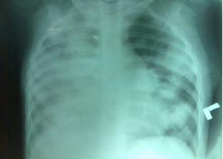
Previously normal 8 years old child, fell from the terrace of first floor with face down into a pool of stagnant water. But for the pain over the legs she had no other complaints on the day of injury. Child was admitted at our institute on the 5th day of injury with breathlessness, chest pain and fever. She was febrile, with a respiratory rate of 60/min, heart rate of 130/min, blood pressure was 100/60mm Hg, capillary refill time was less than 2 seconds. Oxygen saturation was 95% with room air. Examination of the respiratory system revealed bilateral crepitations. Liver span measured 9 cm. Examination of other systems were normal. White blood cell count was 8000cells/cumm (polymorphs 52% lymphocytes 43%, eosinophils 5%). ESR was 30mm in 60 minutes. Liver enzymes, urea, creatinine, electrolytes were within normal limits. Chest X ray showed bilateral infiltrates consistent with aspiration. Ultrasonogram abdomen revealed hepatomegaly, bilateral pleural fluid, free fluid abdomen, increased cortical echoes of both kidneys. C reactive protein (CRP) was positive. Child was started on Injection cefotaxime and amikacin. Child continued to be febrile and tachypneic after 10 days of IV antibiotics. Mantoux test, gastric aspirate for acid fast bacilli and family screening for tuberculosis was negative. Repeat hemogram on the tenth day showed total count of 19,000 cells/cumm (polymorphs 75%, lymphocytes 13%, eosinophils 12%). Chest X-ray showed nodular opacities (figure 1). HRCT chest showed extensive nodular infiltrates. In view of clinical deterioration, extensive nodular pulmonary infiltrates and literature evidence (1) suggesting sedosporium and aspergillus as organisms to be considered in submersion victims as cause for pneumonia, pending fungal culture reports child was started on voriconazole. Child underwent bronchoscopy and bronchoalveolar lavage (BAL) was done which did not grow any organism. Sputum was very scanty and induced sputum after nebulisation showed a few fungal elements. Repeat bronchoscopy was done and bal wet mount was suggestive of fungal elements and nocardia.
BAL fluid was inoculated on blood agar, MacConkey, SDA and thioglycollate broth. After incubation for 24 hrs at 37ºC, blood agar plate showed non hemolytic small, smooth, convex, round, adherent white colonies, and similar colonies were seen in SDA after 48 hours incubation. In the thioglycollate broth, small dull white sand like grains were seen. Direct smear of gram staining showed gram positive filamentous, beaded bacilli with many pus cells. Filamentous, beaded acid fast bacilli were seen in modified acid fast staining. The organism was identified as Nocadia asteroides by hydrolysis of casein, urea and gelatin, and fermentation of glucose, galactose, inositol, ramnose, adonitol, xylose, trehalose and citrate. It was found to be sensitive to amikacin, gentamicin, ciprofloxacin, cotrimoxazole, imipenem and linezolid. SDA also grew buff coloured colonies, which were brown on the reverse. Microscopic examination showed conidial heads with biseriate phialides and smooth walled conidia characteristic of Aspergillus terreus.
The diagnosis was invasive pulmonary aspergillosis with nocardiosis. CT brain was normal with no evidence of brain abscess. Retrovirus screening was negative. Nitrozolium blue test done twice during hospitalization was not suggestive of chronic granulomatous disease. Her immunoglobulin profile was normal. Child was started on oral cotrimoxazole to cover nocardia. With persistent fever, ciprofloxacin was added. Cultures from the water collected from the site where the child fell revealed both aspergillus and nocardia. Repeat x ray chest done after 5th week of hospitalization showed clearance of the nodular opacities and the child was discharged on oral voriconazole, cotrimoxazole and ciprofloxacin. Child was followed up after 6 weeks of voriconazole, her liver enzymes were normal. Repeat X -ray showed residual opacities of the right lung at 8 weeks and the child was asymptomatic. ciprofloxacin and cotrimoxazole were continued for 6 months and the child had complete resolution of the opacities in the chest X-ray.
Fig 1 X-ray chest showing the nodular opacities
Though uncommon, nocardia and aspergillus are known to have caused invasive fungal infections in immunocompetent children. Invasive fungal infection has been reported in submersion injury. The commonly reported fungal organisms are Scedosporium apiospermum and aspergillus. (1) Co-infections of aspergillus and nocardia following drowning or otherwise has been described in literature. (2) Large inoculum of organisms entering the lungs directly during submersion injury could have caused this infection. Underlying lung injury due to submersion can predispose for the proliferation of the organisms. Submersion victims may present with typical bacterial organism or fungal or can be polymicrobial. The radiological features described in literature in pulmonary aspergillosis are not commonly seen in children. HRCT findings may not always show the halo sign or the air crescent sign or cavitating lesion. In any child with submersion injury, clinical deterioration with pulmonary involvement should prompt early use of appropriate antifungal therapy. Awaiting the cultures may delay the diagnosis and can result in increasing mortality (3).
Invasive aspergillosis carries a high mortality. Galactomannan(GM), which is a major aspergillus cell wall antigen released during invasive infection can be a useful surrogate marker for aspergillus infection in the serum and bal. The latter has a better sensitivity in comparison to serum GM levels (4). The serum GM test may not be easily available for earlier identification. Pulmonary nocardiosis though common in immunocompromised, has been unusually encountered in immunocompetent individuals (5). Pulmonary nocardiosis usually manifests as persistent nodular infiltrates. Pulmonary involvement is the most common, followed by the brain. All children with invasive pulmonary nocardiosis need to be evaluated for brain abscesses even if asymptomatic. Pulmonary nocardiosis tends to relapse and needs to be followed up even after complete recovery. Mortality in nocardial infections is > 50% despite appropriate therapy.
Thus we conclude that persistent fever with tachypnea, nodular infiltrates in the chest x-ray despite antibiotic coverage following submersion injury should make one suspect unusual organisms and an earlier diagnosis and therapy may be life saving.
Financial support: none
Conflicting interest: none
| | | | Compliance with Ethical Standards | | Funding None | | | | Conflict of Interest None | | |
- Katragkou A, Dotis J, Kotsiou M, Tamiolaki M, Roilides E. Scedosporium apiospermum infection after near-drowning. Mycoses. 2007; 50: 412-421. [CrossRef]
- Leroy P, Smismans A, Seute T. Invasive pulmonary and central nervous system aspergillosis after near-drowning of a child: case report and review of the literature. Pediatrics. 2006; 118: e509-13.
- Ender PT, Dolan MJ. Pneumonia associated with near-drowning. Clin Infect Dis. 1997; 25: 896-907. [CrossRef]
- Danpornprasert P, Foongladda S, Tscheikuna J. Impact of bronchoalveolar lavage galactomannan on the outcome of patients at risk for invasive pulmonary aspergillosis. J Med Assoc Thai. 2010; 93 Suppl 1: S86-93.
- Dias M, Antony B, Pinto H. Spectrum of Nocardiosis -A Report of Three Cases. Journal of Clinical and Diagnostic Research [serial online] 2009 August [cited: 2011 Jun 8]; 3:1682-1684. Available from http://www.jcdr.net/back_issues.asp?issn=0973-709x&year=2009&month=August&volume=3&issue=4&page=1682-1684&id=557.
DOI: https://doi.org/10.7199/ped.oncall.2012.17
|
| Cite this article as: | | Poovazhagi V, S E, Menon T, V N k, Chamundeeswari, Meeran M. Invasive aspergillosis and nocardial pneumonia in an immunocompetent child. Pediatr Oncall J. 2012;9: 20-22. doi: 10.7199/ped.oncall.2012.17 |
|