Ana Filipa de Abreu Paixão1, Luísa Castello-Branco Ribeiro1, Ana Catarina Amorim1, Ana Tavares2
1Department of Pediatrics, Unidade Local de Saúde Amadora-Sintra, Amadora, Portugal, 2Department of Radiology, Unidade Local de Saúde Amadora-Sintra, Amadora, Portugal
Address for Correspondence: Ana Filipa de Abreu Paixão, Hospital Prof. Doutor Fernando Fonseca E.P.E . IC 19, 2720-276 Amadora, Portugal.
Email: anafapaixao@gmail.com
|
Question :A previously healthy 3-year-old female child vaccinated with PCV-13 vaccine presented with a 10-day history of cough and a 5-day history of fever and dyspnea. Chest radiograph and ultrasound showed extensive right lung pneumonia, with a 2 cm pleural effusion and blood analysis revealed elevation of inflammatory markers. She was started on intravenous ampicillin and clindamycin and thoracentesis fluid was suggestive of empyema, with subsequent identification of Streptococcus pneumoniae serotype 3. Antibiotic therapy was changed to penicillin. Per protocol, she received six doses of intrapleural urokinase and the chest tube was removed on day 7, after 48H of no drainage.
Since admission, the patient continued to experience intermittent fever. Chest radiography on day six (Figure 1) and thoracic computed tomography (CT) scan on day eight (Figure 2) are shown below.
Figure 1. Chest radiograph showing a large round hypotransparency occupying almost the entire right hemithorax, and a hypotransparency in the lower third of the right pulmonary field, associated with blunting of the ipsilateral costophrenic angle. 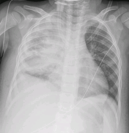 Figure 2.
 Chest CT scan showing a gas containing fluid collection (9x8x6 cm) of the right hemithorax. 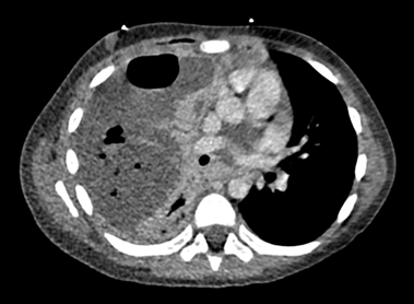 What is the most likely diagnosis?
|
Discussion :
The chest CT scan revealed a large, poorly defined, rounded, gas containing fluid collection (9x8x6 cm) that occupies most of the right hemithorax. The mass is associated with significant atelectasis of the inferior lobe. The severe distortion of lung architecture makes the differential diagnosis between empyema and lung abscess very challenging. While abscesses typically manifest as round lesions with thick well-defined margins, empyemas have a typical lenticular shape, forming an obtuse angle with the chest wall, although they may also present as rounded. Differential diagnosis between these two entities is critical to proper management, since thoracocentesis is typically required for empyemas, but not for lung abscesses. 1 Consulting experienced radiologists and performing serial chest imaging exams is key for diagnosis. Serial chest ultrasounds should start at admission to monitor lesion progression. Consistency and accuracy in interpretation can be ensured when ultrasounds are performed by the same radiologist.
In this patient, serial chest ultrasounds confirmed the presence of necrotizing pneumonia, lung abscess and empyema. Thoracic radiography from day 21 is also shown below (Figure 3). After multidisciplinary discussion, a conservative approach was adopted and the patient completed an eight-week course of antibiotic therapy, resulting in full recovery.
Figure 3. Chest radiograph (day 21) showed extensive right lung necrotizing pneumoniae, lung abscess, empyema and pneumothorax. 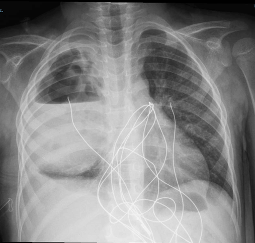 | References : | - Hassan M, Asciak R, Rizk R, Shaarawy H, Gleeson FV, Rahman NM. Lung abscess or empyema? Taking a closer look. Chest Clin. 2018 Mar 26. doi:10.1136/thoraxjnl-2018-211604.
|
|
| Correct Answers : |  100% 100% |
Last Shown : Dec 2025
|