Necrotizing Enterocolitis (nec)
Ira Shah
Consultant Pediatrician, B.J.Wadia Hospital for Children, Mumbai, India
First Created: 01/07/2004
Show details
Introduction
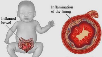
NEC is a syndrome of acute intestinal necrosis. The etiology is unknown and pathogenesis is complex and multifactorial. It is the most common serious surgical disorder among infants in the NICU & a significant cause of neonatal morbidity and mortality. It is seen in 2.5% of all NICU admissions and has an overall mortality rate of 30-40% with mortality increasing to more than 80% in neonates less than 1 kg.
Pathology: Common sites are the terminal ileum and ascending colon.
Predisposing Factors
- Prematurity: Seen commonly at a mean gestational age of 30-32 wks. (AGA). It is the single greatest risk factor. Incidence of NEC increases as gestational age decreases.
- In Term infants & near terms: Conditions predisposing to decreased gastrointestinal oxygen delivery
- Perinatal asphyxia (lower 1 minute apgar score)
- Lower cord PH (acidosis)
- Polycythemia
- RDS/apnea
- Shock
- Umbilical artery catheterization
- Early or large volume nasogastric feeding
- Congenital heart disease
Pathogenesis
- Role of immature Gastrointestinal host defenses
- Role of infectious agents & bacterial toxins
- Role of inflammatory mediators
- Role of oxygen radicals & ischemia reperfusion injury
Clinical Staging Of NEC
| Stage |
Clinical features |
X-ray |
Survival (%) |
|
|
- Mild abdominal distension
- Stasis
- Vomiting
- Poor feeding
|
|
100% |
| - Marked abdominal distension
- GI bleeding
| - Definite ileus
- Pneumatosis intestinalis
|
95% |
| - DIC
- Shock
- Sclerema
- Brownish Peritoneal aspirate
| - Fixed dilated loop of intestine
- Portal vein gas
- Pneumoperitoneum
|
50% |
Clinical Features
- Symptoms may appear within 96 hrs after initiation of feeds
- Majority of cases occur within first 10 days of life
- Onset may be insidious/explosive/delayed
- Earliest signs: abdominal distension, retention of milk in a sick-looking LBW infant.
- Clinical triad: Abdominal. Distension + GI bleeding + Pneumatosis intestinalis
- Signs of functional Intestinal obstruction: Abdominal distension, progressive decreased peristalsis, bilious vomiting, hematemesis and blood in stools
- Signs of peritonitis & perforation: Ascites, erythema & edema of abdominal wall, localized mass or rigidity
- Systemic signs respiratory distress, apnea, bradycardia, lethargy, thermal instability, irritability, poor feeding, hypotension (shock), oliguria, bleeding diathesis, sclerema
Investigations
- Serial X-rays of abdomen: Fixed bowel loop, appearance of mass, pneumatosis intestinalis, portal or hepatic venous air, pneumoperitoneum
- Stool examination: Occult blood, reducing substances for unabsorbed lactose, culture for aerobic/anaerobic bacteria
- Blood: Electrolytes, hematocrit, coagulation status, culture, ABG. Watch for triad of thrombocytopenia, severe refractory hyponatremia and acidosis.
- USG: Micro bubbles of gas in portal vein
- Hydrogen breath test
Management
| Medical |
Surgical |
Respiratory:
- Supplement O2
- Mechanical ventilatory support
CVS:
- Fresh frozen plasma
- Low doses dopamine
Metabolic:
Nutrition:
- Stop oral feedings (7-14 days)
- TPN instituted (90-110 cal/kg/d)
Antibiotics- For 7-14 days. Broad spectrum
Hematological:
- Platelet transfusions
- Packed RBCs
- Vitamin K
CNS:
- Treat IVH, meningitis, seizures
Renal:
|
Indications:
- Bowel perforation
- Full thickness necrosis of bowel wall as evidenced by dilated loop of intestine unchanged in position > 24 hrs.
- Peritonitis. Aspiration of brown colored fluid is indicative of intestinal gangrene.
Surgical treatment:
Excision of necrotic area & end-to-end anastomosis |
Differential Diagnosis
- Pneumonia & sepsis
- Surgical abdominal catastrophes
- Malrotation with obstruction
- Malrotation with midgut volvulus
- Intusussception
- Ulcer
- Gastric perforation
- Mesenteric vessel thrombosis
- Infections enterocolitis with diarrhea
- Inherited metabolic disease
- Feeding intolerance
- Systemic candidiasis
Prevention
Corticosteroids - Prenatal/Postnatal
- Lower incidence of NEC in mothers of preterm who received dexamethasone antenatally.
Feeding regimen
- Breast feeding
- Acidified feeds
- Small iso-osmolar feeds with gradual increase in feeds
Oral Immunoglobulins (IgA & IgG)
- Enhances intestinal immune defenses
Reduce incidence of:
- Preterm delivery
- Prevent predisposing factors
Prognosis
Recurrent NEC is 4%
Sequelae
- Strictures (20%) most common in large bowel
- Enteric fistulas
- Short bowel syndrome (following surgery)
- Malabsorption & chronic diarrhea
- Dumping syndromes: loss of terminal ileum
- Fluids electrolyte losses (with ileostomy)
- Parenteral nutrition associated hepatic disease
- Developmental delay
 Ira Shah
Necrotizing Enterocolitis (NEC)
https://www.pediatriconcall.com/show_article/default.aspx?main_cat=neonatology&sub_cat=necrotizing-enterocolitis-nec&url=necrotizing-enterocolitis-nec-introduction
2004-01-07
2004-01-07
Ira Shah
Necrotizing Enterocolitis (NEC)
https://www.pediatriconcall.com/show_article/default.aspx?main_cat=neonatology&sub_cat=necrotizing-enterocolitis-nec&url=necrotizing-enterocolitis-nec-introduction
2004-01-07
2004-01-07
×
Contributor Information and Disclosures
Ira Shah
Consultant Pediatrician, B.J.Wadia Hospital for Children, Mumbai, India
First Created: 01/07/2004